Fraud Blocker Skip to main content

Malignancy Collection (page 4)

"Malignancy: Unveiling the Battle Within" In this captivating journey through the realm of malignancy

Background imageMalignancy Collection: Colon cancer, endoscopic view C016 / 8307

Colon cancer, endoscopic view C016 / 8307
Colon cancer. Endoscopic view of a malignant (cancerous) adenocarcinoma (lower right) in a patients ascending colon (large intestine)

Background imageMalignancy Collection: Blood vessel formation, artwork

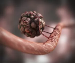
Blood vessel formation, artwork
Blood vessel formation. Artwork showing malignant (cancerous) tumour cells promoting the formation of new blood vessels, a process known as angiogenesis

Background imageMalignancy Collection: Dividing cancer cell, SEM C014 / 0361

Dividing cancer cell, SEM C014 / 0361
Dividing cancer cell. Coloured scanning electron micrograph (SEM) of a colorectal cancer cell undergoing mitosis (nuclear division) and splitting into two daughter cells (left and right)

Background imageMalignancy Collection: Dividing brain cancer cells, SEM C014 / 0354

Dividing brain cancer cells, SEM C014 / 0354
Dividing brain cancer cells. Coloured scanning electron micrograph (SEM) of a cancerous astrocyte brain cell that has just undergone cytokinesis (cell division)
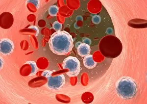
Background imageMalignancy Collection: Dividing bone cancer cell, SEM

Dividing bone cancer cell, SEM
SEM Dividing Osteosarcoma cells Osteosarcoma is an aggressive malignant neoplasm arising from primitive transformed cells of mesenchymal origin (and thus a sarcoma)

Background imageMalignancy Collection: Cervical cancer, light micrograph C015 / 7000

Cervical cancer, light micrograph C015 / 7000
Cervical cancer. Light micrograph of a section through tissue from the cervix (neck of the womb) showing intraepithelial carcinoma (centre), a form of cancer
Background imageMalignancy Collection: Cervical cancer, light micrograph C015 / 6999

Cervical cancer, light micrograph C015 / 6999
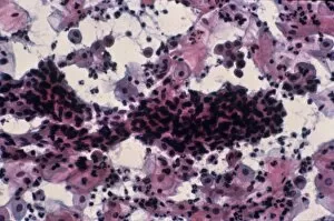
Cervical cancer. Light micrograph of a section through tissue from the cervix (neck of the womb) showing intraepithelial carcinoma (across centre), a form of cancer

Background imageMalignancy Collection: Cervical cancer, light micrograph C015 / 6998

Cervical cancer, light micrograph C015 / 6998
Cervical cancer. Light micrograph of a section through tissue from the cervix (neck of the womb) showing intraepithelial carcinoma (dark areas), a form of cancer

Background imageMalignancy Collection: Cervical cancer, light micrograph C015 / 6997

Cervical cancer, light micrograph C015 / 6997
Cervical cancer. Light micrograph of a section through tissue from the cervix (neck of the womb) showing intraepithelial carcinoma (across centre), a form of cancer

Background imageMalignancy Collection: Cervical smear showing endometrial cancer C015 / 6750

Cervical smear showing endometrial cancer C015 / 6750
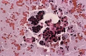
Cervical smear showing endometrial cancer. Light micrograph of cells from a cervical smear test showing endometrial sarcoma. This is a cancer of the lining of the womb, the endometrium

Background imageMalignancy Collection: Cancer cell, SEM C014 / 0628

Cancer cell, SEM C014 / 0628
Cancer cell. Coloured scanning electron micrograph (SEM) of a carcinoma cancer cell (pink) migrating (metastasising) along a layer of normal epithelial cells (purple)

Background imageMalignancy Collection: Cancer cell, SEM C014 / 0627

Cancer cell, SEM C014 / 0627
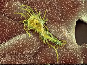
Cancer cell. Coloured scanning electron micrograph (SEM) of a carcinoma cancer cell (yellow) migrating (metastasising) along a layer of normal epithelial cells (brown)

Background imageMalignancy Collection: Throat cancer, artwork C013 / 5939

Throat cancer, artwork C013 / 5939
Throat cancer. Cutaway computer artwork of a persons head, showing cancer (yellow) in the pharynx (centre) and oesophagus (lower centre)

Background imageMalignancy Collection: Endometrial cancer, light micrograph C015 / 6061

Endometrial cancer, light micrograph C015 / 6061
Endometrial cancer. Light micrograph of a section through a tumour in the endometrium, the lining of the uterus (womb). Endometrial cancer is the third most common cause of gynaecological cancer

Background imageMalignancy Collection: Bone cancer cell, SEM

Bone cancer cell, SEM
Bone cancer cell. Coloured scanning electron micrograph of a fibrosarcoma (fibroblastic sarcoma) cell, showing the numerous filopodia (hair-like) covering its surface

Background imageMalignancy Collection: HeLa cell dying, SEM C017 / 8304

HeLa cell dying, SEM C017 / 8304
HeLa cell dying, coloured scanning electron micrograph (SEM). The cell in the foreground is undergoing apoptosis, or programmed cell death. Apoptosis occurs when a cell becomes old or damaged

Background imageMalignancy Collection: Mesothelioma lung cancer, CT scan

Mesothelioma lung cancer, CT scan
Mesothelioma lung cancer. Computed tomography (CT) scan of the lungs of a 69 year old patient with mesothelioma, a type of lung cancer

Background imageMalignancy Collection: Lung cancer, CT and PET scans

Lung cancer, CT and PET scans
Lung cancer. Combined computed tomography (CT) and positron emission tomography (PET) scans of the lungs of an 84 year old woman with a malignant (cancerous) tumour (bright patch)

Background imageMalignancy Collection: Lung cancer, X-ray C017 / 7964

Lung cancer, X-ray C017 / 7964
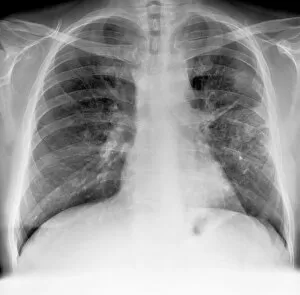
Lung cancer. X-ray if the chest of a patient with malignant (cancerous) tumours in middle of the left lung (seen at right). The main cause of lung cancer is cigarette smoke

Background imageMalignancy Collection: Chronic myeloid leukaemia, micrograph C015 / 6227

Chronic myeloid leukaemia, micrograph C015 / 6227
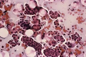
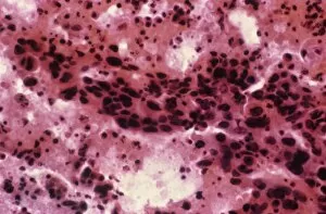
Chronic myeloid leukaemia. Light micrograph of bone marrow from a patient with chronic myeloid leukaemia (CML). This cancer affects the myeloid tissue (bone marrow)

Background imageMalignancy Collection: Acute myeloid leukaemia C015 / 6225

Acute myeloid leukaemia C015 / 6225
Acute myeloid leukaemia. Light micrograph of bone marrow from a patient with acute myeloid leukaemia (AML). This cancer affects the myeloid tissue (bone marrow)

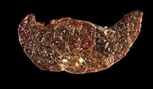
Background imageMalignancy Collection: Spleen cancer, gross specimen C015 / 6421

Spleen cancer, gross specimen C015 / 6421
Spleen cancer. Gross specimen of a spleen showing a large lymphosarcoma, or non-Hodgkins lymphoma, a cancer of lymphoid tissue. The lymphatic system is a major part of the immune system

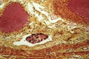
Background imageMalignancy Collection: Tumour embolism, light micrograph C015 / 6397

Tumour embolism, light micrograph C015 / 6397
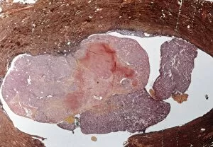
Tumour embolism. Light micrograph of a section through an embolism (blood clot) caused by cells from a cervical cancer. The cells have left the primary tumour and are metastasising (spreading)

Background imageMalignancy Collection: Mesothelioma lung cancer, X-ray C017 / 7776

Mesothelioma lung cancer, X-ray C017 / 7776
Mesothelioma lung cancer, X-ray of the chest of a 84 year old male patient with advanced mesothelioma, a type of lung cancer

Background imageMalignancy Collection: Angiosarcoma of the spleen C015 / 6398

Angiosarcoma of the spleen C015 / 6398
Angiosarcoma. Gross specimen of a sectioned spleen affected by angiosarcoma. Malignant (cancerous) tumours (yellow) can be seen at bottom centre

Background imageMalignancy Collection: Secondary pancreatic cancer, ultrasound C017 / 7765

Secondary pancreatic cancer, ultrasound C017 / 7765
Secondary pancreatic cancer. Ultrasound scan of a 63 year old male patients pancreas showing metastatic (secondary) tumours. The cancer has metastasised (spread) from a primary cancer

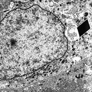
Background imageMalignancy Collection: Alveolar soft part sarcoma, TEM C016 / 5826

Alveolar soft part sarcoma, TEM C016 / 5826
Alveolar soft part sarcoma. Transmission electron micrograph (TEM) of a section through an alveolar soft part sarcoma (ASPS) showing a characteristic rhomboid crystal (black, centre)

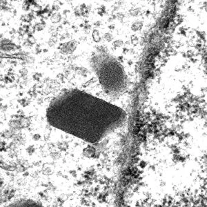
Background imageMalignancy Collection: Alveolar soft part sarcoma, TEM C016 / 5816

Alveolar soft part sarcoma, TEM C016 / 5816
Alveolar soft part sarcoma. Transmission electron micrograph (TEM) of a section through an alveolar soft part sarcoma (ASPS) showing a characteristic rhomboid crystal (black)

Background imageMalignancy Collection: Brain cancer, MRI scan C016 / 4438

Brain cancer, MRI scan C016 / 4438
Brain cancer. Coloured coronal magnetic resonance imaging (MRI) scan through the head of a 48 year old male patient with a glioblastoma (red)

Background imageMalignancy Collection: Brain cancer, MRI scan C016 / 4436

Brain cancer, MRI scan C016 / 4436
Brain cancer. Coloured coronal magnetic resonance imaging (MRI) scan through the head of a 48 year old male patient with a glioblastoma (centre right)

Background imageMalignancy Collection: Brain cancer, MRI scan C016 / 4437

Brain cancer, MRI scan C016 / 4437
Brain cancer. Coloured coronal magnetic resonance imaging (MRI) scan through the head of a 48 year old male patient with a glioblastoma (orange)

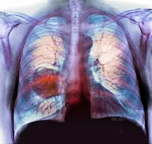
Background imageMalignancy Collection: Lung cancer, X-ray C016 / 4435

Lung cancer, X-ray C016 / 4435
Lung cancer. Coloured chest X-ray of a 64 year old male patient with lung cancer. The tumour is seen as the red mass in the right lung (bright area at left)

Background imageMalignancy Collection: Lung cancer, X-ray C016 / 4432

Lung cancer, X-ray C016 / 4432
Lung cancer. Coloured chest X-ray of a 64 year old male patient with lung cancer. The tumour is seen as the orange mass in the right lung (dark area at left)

Background imageMalignancy Collection: Lung cancer, X-ray C016 / 4433

Lung cancer, X-ray C016 / 4433
Lung cancer. Coloured chest X-ray of a 64 year old male patient with lung cancer. The tumour is seen as the orange mass in the right lung (bright area at left)

Background imageMalignancy Collection: Lung cancer cell division, artwork C013 / 4630

Lung cancer cell division, artwork C013 / 4630
Lung cancer cell division. Computer artwork of a lung cancer cell during cell division (cytokinesis). The two daughter cells remain temporarily joined by a cytoplasmic bridge (centre)

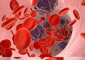
Background imageMalignancy Collection: Blood cancer, conceptual image C013 / 4618

Blood cancer, conceptual image C013 / 4618
Blood cancer, conceptual image. Computer artwork of the interior of a blood vessel, showing cancer cells (dark) amongst normal red blood cells (red)

Background imageMalignancy Collection: Blood cancer, conceptual image C013 / 4614

Blood cancer, conceptual image C013 / 4614
Blood cancer, conceptual image. Computer artwork of the interior of a blood vessel, showing cancer cells (white) amongst normal red blood cells (red)

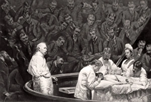
Background imageMalignancy Collection: Agnews surgical clinic, 1889

Agnews surgical clinic, 1889
Agnews surgical clinic. At left, holding a scalpel, is US surgeon David Hayes Agnew (1818-1892), supervising a partial breast removal operation (mastectomy)

Background imageMalignancy Collection: Prostate cancer cell, SEM C013 / 5012

Prostate cancer cell, SEM C013 / 5012
Prostate cancer cell, coloured scanning electron micrograph (SEM). The uneven surface of this cell is typical of cancer cells

Background imageMalignancy Collection: Prostate cancer cell, SEM C013 / 5011

Prostate cancer cell, SEM C013 / 5011
Prostate cancer cell, coloured scanning electron micrograph (SEM). The uneven surface of this cell is typical of cancer cells

Background imageMalignancy Collection: T lymphocytes and cancer cells, SEM

T lymphocytes and cancer cells, SEM
T lymphocytes and cancer cells. Coloured scanning electron micrograph (SEM) of T lymphocyte cells (purple) attached to cancer cells

Background imageMalignancy Collection: Coloured SEM of lymphocyte attacking cancer cell

Coloured SEM of lymphocyte attacking cancer cell
ARRAY(0xd68c618) Commercial use requires clearance

Background imageMalignancy Collection: Coloured SEM of a leukaemic white blood cell

Coloured SEM of a leukaemic white blood cell

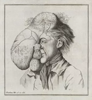
Background imageMalignancy Collection: Large tumour of the head, 18th century

Large tumour of the head, 18th century


All Professionally Made to Order for Quick Shipping

"Malignancy: Unveiling the Battle Within" In this captivating journey through the realm of malignancy, we witness the relentless struggle between T lymphocytes and cancer cells. A mesmerizing SEM image (C001 / 1679) reveals a colorful spectacle as lymphocytes valiantly attack a cancer cell, embodying hope in the face of adversity. Moving deeper into this intricate world, an ovarian cancer light micrograph (C015 / 7103) unveils the hidden intricacies of this disease. The delicate brushstrokes of an oil painting depicting breast cancer (C017 / 1273) evoke both beauty and pain, reminding us that art can capture even the most complex emotions. Venturing into the depths of our minds, brain tumors come to life through fMRI and tractography scans (C017 / 7102). These images showcase not only their physical presence but also their impact on neural pathways. DTI modeling (C017 / 7060) further unravels these enigmatic growths while three-dimensional CT scans (C016 / 6414) provide a comprehensive view from all angles. The battle against malignancy extends beyond just one organ or system. Throat cancer X-rays and secondary lung cancers X-ray serve as stark reminders that no part is immune to its reach. An ultrasound scan exposes secondary liver cancer's insidious invasion (C017/7764), urging us to remain vigilant in our fight against this formidable foe. A cross-section diagram portrays a vivid snapshot of a malignant tumor's complexity - calcium deposits, blood vessels, tumorous outgrowths - all entwined with nerve fibers amidst ulcerated areas and bleeding wounds. This visual representation serves as a reminder that behind every diagnosis lies an intricate web affecting multiple facets of our being. As science progresses hand-in-hand with compassion, we delve deeper into understanding malignancies' secrets.