Fraud Blocker Skip to main content

Macromolecule Collection (#9)

Macromolecules, the building blocks of life, are at the forefront of scientific innovation

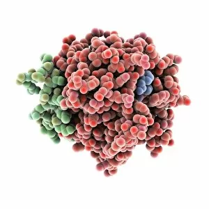
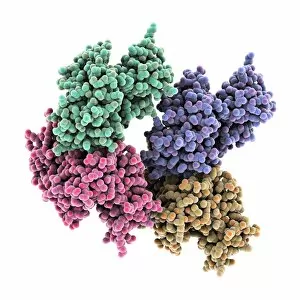
Background imageMacromolecule Collection: Thrombin protein, molecular model C015 / 7074

Thrombin protein, molecular model C015 / 7074
Thrombin protein, molecular model. Thrombin is an enzyme involved in the blood coagulation (clotting) process. It converts fibrinogen (a soluble plasma glycoprotein synthesised in the liver)

Background imageMacromolecule Collection: Simian virus (SV40) large T antigen C015 / 7070

Simian virus (SV40) large T antigen C015 / 7070
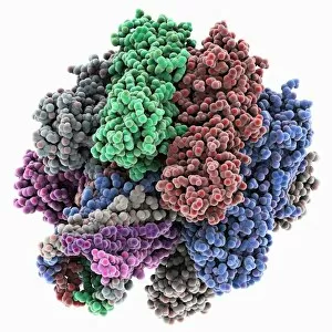
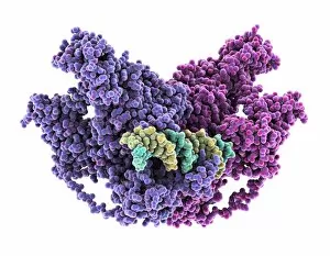
Simian virus (SV40) large T antigen, molecular model. This antigen is from the simian vacuolating virus 40 (SV40). Large T antigens play a role in regulating the viral life cycle of

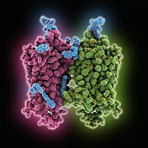
Background imageMacromolecule Collection: Bacterial alpha-hemolysin toxin C015 / 7067

Bacterial alpha-hemolysin toxin C015 / 7067
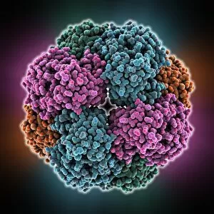
Bacterial alpha-hemolysin toxin, molecular model. This toxin is secreted by the bacterium Staphylococcus aureus. It is an example of a pore-forming toxin

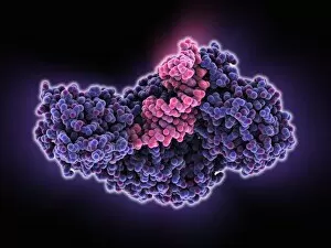
Background imageMacromolecule Collection: Sir3 gene silencer acting on DNA C015 / 7062

Sir3 gene silencer acting on DNA C015 / 7062
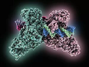
Sir3 gene silencer acting on DNA, molecular model. Sir3 (purple and grey) is acting on a circular strand of DNA (deoxyribonucleic acid, red)

Background imageMacromolecule Collection: Sir3 gene silencer acting on DNA C015 / 7061

Sir3 gene silencer acting on DNA C015 / 7061
Sir3 gene silencer acting on DNA

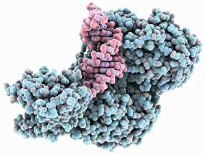
Background imageMacromolecule Collection: TFAM transcription factor bound to DNA C015 / 7060

TFAM transcription factor bound to DNA C015 / 7060
TFAM transcription factor bound to DNA, molecular model. Human mitochondrial transcription factor A (TFAM, pink) bound to a strand of DNA (deoxyribonucleic acid, orange and green)

Background imageMacromolecule Collection: Peroxiredoxin 4 antioxidant enzyme C015 / 7022

Peroxiredoxin 4 antioxidant enzyme C015 / 7022
Peroxiredoxin 4 antioxidant enzyme, molecular model. This enzyme, also called peroxiredoxin IV (PrxIV), plays a catalytic role in cell metabolism on the endoplasmic reticulum

Background imageMacromolecule Collection: Mandelate racemase enzyme C015 / 7021

Mandelate racemase enzyme C015 / 7021
Mandelate racemase enzyme, molecular model. This bacterial enzyme is an example of a muconate lactonizing enzyme, and plays a key role in the metabolism of soil and aquatic bacteria

Background imageMacromolecule Collection: Mandelate racemase enzyme C015 / 7020

Mandelate racemase enzyme C015 / 7020
Mandelate racemase enzyme, molecular model. This bacterial enzyme is an example of a muconate lactonizing enzyme, and plays a key role in the metabolism of soil and aquatic bacteria

Background imageMacromolecule Collection: Nucleoplasmin histone-chaperone protein C015 / 6915

Nucleoplasmin histone-chaperone protein C015 / 6915
Nucleoplasmin histone-chaperone protein, molecular model. This is nucleoplasmin 2 (Npm2), a chaperone that acts on human nucleoplasmin. Chaparone proteins play a key role in aiding protein folding

Background imageMacromolecule Collection: Methyltransferase complexed with DNA C016 / 2033

Methyltransferase complexed with DNA C016 / 2033
Methyltransferase complexed with DNA, molecular model. The strand of DNA (deoxyribonucleic acid, green and blue) is enclosed by DNA methyltransferase 1 (DNMT-1, green and pink)

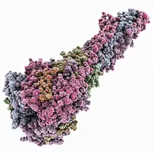
Background imageMacromolecule Collection: Newcastle disease virus protein C015 / 6912

Newcastle disease virus protein C015 / 6912
Newcastle disease virus protein, molecular model. This is a hemagglutinin-neuromidase (HN) protein that enables the virus to bind to and infect host cells

Background imageMacromolecule Collection: Methyltransferase complexed with DNA C016 / 2032

Methyltransferase complexed with DNA C016 / 2032
Methyltransferase complexed with DNA, molecular model. The strand of DNA (deoxyribonucleic acid, green and yellow) is enclosed by DNA methyltransferase 1 (DNMT-1, purple and pink)

Background imageMacromolecule Collection: NpmA methyltransferase C016 / 2031

NpmA methyltransferase C016 / 2031
NpmA methyltransferase, molecular model. Methyltransferase enzymes act to add methyl groups to nucleic acids such as DNA, a process called DNA methylation

Background imageMacromolecule Collection: NpmA methyltransferase C016 / 2030

NpmA methyltransferase C016 / 2030
NpmA methyltransferase, molecular model. Methyltransferase enzymes act to add methyl groups to nucleic acids such as DNA, a process called DNA methylation

Background imageMacromolecule Collection: Notch transcription, molecular model

Notch transcription, molecular model
Notch transcription. Molecular model showing a strand of DNA (deoxyribonucleic acid) being acted upon by various proteins and other molecules

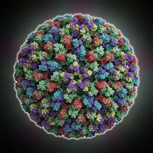
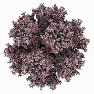
Background imageMacromolecule Collection: Penicillium partitivirus capsid

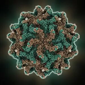
Penicillium partitivirus capsid, molecular model. This is the capsid of the partivirus called Penicillium stoloniferum virus F (PsV-F). This virus infects the fungi that make the drug penicillin

Background imageMacromolecule Collection: Plant agglutinin protein C015 / 6107

Plant agglutinin protein C015 / 6107
Plant agglutinin protein, molecular model. This example, from the hedge-apple (Maclura pomifera), is known as Maclura pomifera agglutinin (MPA). It is a tetrameric plant seed lectin

Background imageMacromolecule Collection: Apoptosome cell death protein C015 / 6108

Apoptosome cell death protein C015 / 6108
Apoptosome cell death protein, molecular model. Apoptosomes are large protein structures formed during programmed cell death (PCD, also called apoptosis)

Background imageMacromolecule Collection: Plant agglutinin protein C015 / 6106

Plant agglutinin protein C015 / 6106
Plant agglutinin protein, molecular model. This example, from the hedge-apple (Maclura pomifera), is known as Maclura pomifera agglutinin (MPA). It is a tetrameric plant seed lectin

Background imageMacromolecule Collection: Echovirus 7 capsid, molecular model

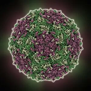
Echovirus 7 capsid, molecular model. Echoviruses are related to the polioviruses. They are usually harmless but can cause serious illness such as encephalitis, meningitis, heart and liver disease

Background imageMacromolecule Collection: Foot-and-mouth disease virus capsid

Foot-and-mouth disease virus capsid, molecular model. This virus, which can be fatal, causes foot-and-mouth disease in cloven-hooved animals

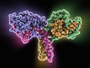
Background imageMacromolecule Collection: C3-degrading proteinase enzyme C016 / 1363

C3-degrading proteinase enzyme C016 / 1363
C3-degrading proteinase enzyme. Molecular model of a proteinase enzyme from the TIGR4 strain of the bacterium Streptococcus pneumoniae
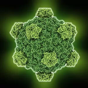
Background imageMacromolecule Collection: Human interleukin-28B C016 / 1364

Human interleukin-28B C016 / 1364
Human interleukin-28B (IL-28B), molecular model. Interleukins a group of secreted proteins and signalling molecules that play a range of roles in cellular communication

Background imageMacromolecule Collection: Poliovirus type 1 capsid, molecular model

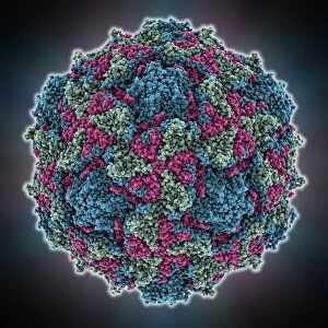
Poliovirus type 1 capsid, molecular model. This enterovirus causes poliomyelitis (polio) in humans, which affects the nervous system, sometimes leading to paralysis

Background imageMacromolecule Collection: Human interleukin-28B C016 / 1365

Human interleukin-28B C016 / 1365
Human interleukin-28B (IL-28B), molecular model. Interleukins a group of secreted proteins and signalling molecules that play a range of roles in cellular communication

Background imageMacromolecule Collection: Beta secretase inhibitor, molecular model

Beta secretase inhibitor, molecular model. The inhibitor molecule (centre, also shown in C015/1977) is bound to the beta secretase enzyme (partially seen)

Background imageMacromolecule Collection: IBDV subviral particle, molecular model

IBDV subviral particle, molecular model. IBDV (infectious bursal disease virus) is an avian virus that infects the bursa of Fabricius (specialised bird immune organ) in young chickens

Background imageMacromolecule Collection: C3-degrading proteinase enzyme C016 / 1362

C3-degrading proteinase enzyme C016 / 1362
C3-degrading proteinase enzyme. Molecular model of a proteinase enzyme from the TIGR4 strain of the bacterium Streptococcus pneumoniae

Background imageMacromolecule Collection: Cholera virulence regulator protein C015 / 6727

Cholera virulence regulator protein C015 / 6727
Cholera virulence regulator protein, molecular model. This protein is AphB, a virulence gene activator from the Vibrio cholerae bacterium

Background imageMacromolecule Collection: Bluetongue virus capsid

Bluetongue virus capsid
Bluetongue virus (BTV) capsid, molecular model. BTV is an orbivirus, and is of major economic importance as an insect-borne pathogen of cattle and other ruminants

Background imageMacromolecule Collection: Acyl-CoA hydrolase enzyme C015 / 6724

Acyl-CoA hydrolase enzyme C015 / 6724
Acyl-CoA hydrolase enzyme, molecular model. This is an acyl-CoA thioesterase enzyme. Thioesterases hydrolyze coenzyme A (CoA) esters, producing the free acid and CoA

Background imageMacromolecule Collection: Acyl-CoA hydrolase enzyme C015 / 6725

Acyl-CoA hydrolase enzyme C015 / 6725
Acyl-CoA hydrolase enzyme, molecular model. This is an acyl-CoA thioesterase enzyme. Thioesterases hydrolyze coenzyme A (CoA) esters, producing the free acid and CoA

Background imageMacromolecule Collection: Cholera virulence regulator protein C015 / 6726

Cholera virulence regulator protein C015 / 6726
Cholera virulence regulator protein, molecular model. This protein is AphB, a virulence gene activator from the Vibrio cholerae bacterium

Background imageMacromolecule Collection: Opsin molecule C016 / 0723

Opsin molecule C016 / 0723
Opsin. Molecular model of a ligand-free opsin molecule. Opsins are found in photoreceptor cells (rods and cones) in the retina of the eye

Background imageMacromolecule Collection: Haemoglobin S, molecular model

Haemoglobin S, molecular model
Haemoglobin S. Molecular model of the mutant form of haemoglobin (haemoglobin S) that causes sickle cell anaemia. This is deoxyhaemoglobin S, the molecule in its non-oxygen bound state

Background imageMacromolecule Collection: Alpha 3 bacteriophage capsid

Alpha 3 bacteriophage capsid, molecular model. Bacteriophages are viruses that infect bacteria, with the capsid forming the bacteriophage head. A capsid has subunits called capsomeres

Background imageMacromolecule Collection: Opsin molecule C016 / 0635

Opsin molecule C016 / 0635
Opsin. Molecular model of a ligand-free opsin molecule. Opsins are found in photoreceptor cells (rods and cones) in the retina of the eye

Background imageMacromolecule Collection: Iron-regulatory protein bound to RNA C015 / 6691

Iron-regulatory protein bound to RNA C015 / 6691
Iron-regulatory protein bound to RNA, molecular model. Iron regulatory protein 1 (IRP1, purple) bound to a short strand of RNA (ribonucleic acid, pink) that includes iron-responsive elements (IREs)

Background imageMacromolecule Collection: Iron-regulatory protein bound to RNA C015 / 6690

Iron-regulatory protein bound to RNA C015 / 6690
Iron-regulatory protein bound to RNA, molecular model. Iron regulatory protein 1 (IRP1, blue) bound to a short strand of RNA (ribonucleic acid, pink) that includes iron-responsive elements (IREs)

Background imageMacromolecule Collection: DNA hybrid duplex, molecular model

DNA hybrid duplex, molecular model. This model shows a chimeric junction, where a DNA (deoxyribonucleic acid) strand changes from one form to another

Background imageMacromolecule Collection: RSV virus fusion glycoprotein C015 / 6689

RSV virus fusion glycoprotein C015 / 6689
RSV virus fusion glycoprotein, molecular model. This is a fusion glycoprotein of human respiratory syncytial virus (RSV). This glycoprotein is used by the virus to invade host cells

Background imageMacromolecule Collection: RSV virus fusion glycoprotein C015 / 6688

RSV virus fusion glycoprotein C015 / 6688
RSV virus fusion glycoprotein, molecular model. This is a fusion glycoprotein of human respiratory syncytial virus (RSV). This glycoprotein is used by the virus to invade host cells

Background imageMacromolecule Collection: Adenovirus penton base protein

Adenovirus penton base protein, molecular model. This protein molecule is a subunit called a penton, forming the vertices of the capsid of this adenovirus

Background imageMacromolecule Collection: Aquaporin membrane protein C015 / 5922

Aquaporin membrane protein C015 / 5922
Aquaporin membrane protein, molecular model. Aquaporins are membrane proteins that form channels (lower right) that help water molecules pass in and out of cells

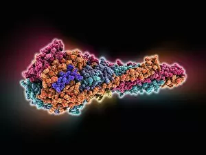
Background imageMacromolecule Collection: Kinesin motor protein dimer C015 / 5921

Kinesin motor protein dimer C015 / 5921
Kinesin motor protein dimer, molecular model. Kinesin is a motor protein that moves along microtubule filaments in cells. It does so by forming a dimer, the heads of which walk along the microtubule

Background imageMacromolecule Collection: Kinesin motor protein dimer C015 / 5920

Kinesin motor protein dimer C015 / 5920
Kinesin motor protein dimer, molecular model. Kinesin is a motor protein that moves along microtubule filaments in cells. It does so by forming a dimer, the heads of which walk along the microtubule

Background imageMacromolecule Collection: Transcription repressor protein and DNA C015 / 5810

Transcription repressor protein and DNA C015 / 5810
Transcription repressor protein and DNA, molecular model. The repressor protein (green) is binding to a strand of DNA (deoxyribonucleic acid, pink and purple)


All Professionally Made to Order for Quick Shipping

Macromolecules, the building blocks of life, are at the forefront of scientific innovation. Nanotube technology has revolutionized various fields, enabling advancements in medicine and electronics. In this captivating computer artwork, we witness the intricate Zinc fingers binding to a DNA strand, showcasing their crucial role in gene regulation. Carbon nanotubes have also emerged as remarkable materials with immense potential. Their unique structure and properties make them ideal for applications ranging from energy storage to drug delivery systems. Computer-generated images depict these carbon nanotubes in all their glory. The SARS coronavirus protein is another macromolecule that has garnered significant attention due to its role in viral infection. Scientists tirelessly study it to develop effective treatments against deadly outbreaks. Computer models allow us to explore complex structures like Bacteriophage phi29—a virus that infects bacteria—providing insights into its mechanisms and aiding in the development of targeted therapies. Simian immunodeficiency virus (SIV), closely related to HIV, poses a global health challenge. Understanding its macromolecular components helps researchers devise strategies for prevention and treatment. Rhodopsin protein molecule captures our imagination with its vital function in vision. Its elegant structure enables light detection and initiates visual signals within our eyes. TFAM transcription factor bound to DNA C015/7059 showcases how macromolecules regulate gene expression by interacting with specific regions on DNA strands—an essential process for cell functioning and development. These glimpses into the world of macromolecules highlight their significance across diverse disciplines—from cutting-edge technologies like nanotube engineering to unraveling infectious diseases or understanding fundamental biological processes. As scientists continue exploring these fascinating molecules, they pave the way for groundbreaking discoveries that shape our future.