
Lung Cancer Collection
In the 19th century, Crown Prince Frederick William of Prussia may have never imagined the devastating impact that lung cancer would have on future generations


All Professionally Made to Order for Quick Shipping
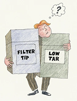
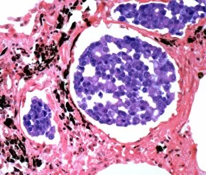
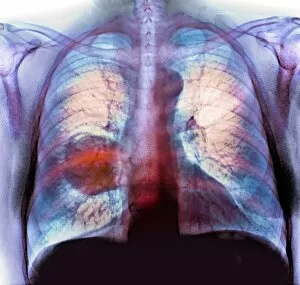
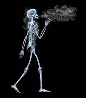
In the 19th century, Crown Prince Frederick William of Prussia may have never imagined the devastating impact that lung cancer would have on future generations. As we look at an illustration of a man with a question mark above his head, holding two oversized filter tip and low tar cigarette packs, it becomes evident that society has long been puzzled by the dangers of smoking. An artist's depiction of mesothelioma in the lungs and abdominal cavity serves as a stark reminder that they are also be caused by exposure to harmful substances like asbestos. The accompanying artworks F007/6159, F007/6161, F007/6160, and F007/6162 further emphasize the link between smoking and this deadly disease. Even female anatomy is not spared from the clutches of lung cancer, as depicted in various artworks. These illustrations highlight how no gender or body is immune to its effects. Artwork F008/0116 portrays lung cancer itself while artwork F008/0214 showcases secondary lung cancer through X-ray imagery. As we reflect upon these visuals, it becomes clear that raising awareness about lung cancer is crucial for prevention and early detection. Let us remember Crown Prince Frederick William's era when contemplating our choices today - choosing health over addiction could spare countless lives from falling victim to this merciless disease.