
Lobes Collection
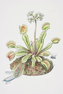
"Lobes: Nature's Traps and Marvels" In the enchanting world of Dionaea muscipula, commonly known as the Venus Fly Trap, lobes take on a whole new meaning


All Professionally Made to Order for Quick Shipping
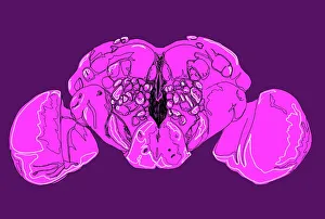
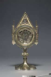
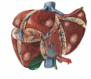
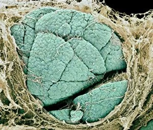
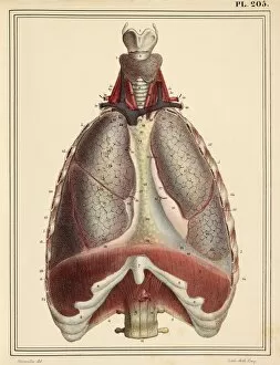
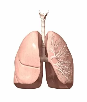
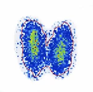
"Lobes: Nature's Traps and Marvels" In the enchanting world of Dionaea muscipula, commonly known as the Venus Fly Trap, lobes take on a whole new meaning. These carnivorous plants possess specialized leaves that form two hinged lobes, ready to snap shut at any moment. Imagine a delicate Dragonfly innocently perched on one of these vibrant flowering plants, unaware of its impending doom caught in the clutches of the lobes. But it's not just insects that fall victim to these captivating lobes; even Houseflies hover nearby, enticed by their irresistible allure. As they cautiously approach, little do they know that within seconds those very same lobes will trap them tightly. Moving away from nature's wonders, let us delve into another realm where lobes play a crucial role – our own bodies. The Basal ganglia is an intricate network deep within our brains composed of different interconnected structures or "lobes. " It regulates movement and plays a vital part in various cognitive functions. Artists have also found inspiration in exploring the concept of lobes. An artwork featuring two Dionaea muscipula showcases one lobe opening wide to catch a fly while the other traps it between closed jaws—a front-row view into nature's cunning mechanisms. Delving further into scientific illustrations, we discover an intricate depiction of a Fruit fly brain with its complex web-like structure beautifully captured through illustration C018 / 0791. This mesmerizing image reminds us how even tiny creatures possess remarkable intricacies hidden within their small frames. Shifting gears once more, we explore SEM images revealing Thyroid gland capillaries and blood vessels—microscopic networks resembling delicate lacework spread throughout this essential organ responsible for regulating metabolism and hormone production. Our journey takes us across continents as we encounter cultural artifacts like libation bowls adorned with embossed decorations shaped like multipetaled rosettes made from precious gold.