Fraud Blocker Skip to main content

Legionella Pneumophila Collection

"Unveiling the Transformation: Legionella pneumophila, from Saul's Conversion to Microscopic Marvels" Witness the remarkable journey of Legionella pneumophila

Background imageLegionella Pneumophila Collection: Saul’s conversion

Saul’s conversion
[url=http://www.istockphoto.com/file_search.php?action=file&lightboxID=11047139][img]http://img-fotki.yandex.ru/get/5809/5232617.2/0_702d2_40b270_orig[/img][/url] Saul is struck by God

Background imageLegionella Pneumophila Collection: Conversion of Saint Paul

Conversion of Saint Paul
" Conversion of Saint Paul, a scene from the bible. Engraving from 1870. Engraving by Gustave Dore, Photo by D Walker."

Background imageLegionella Pneumophila Collection: Defeat of the Cimbri at Vercellae 101 B. C

Defeat of the Cimbri at Vercellae 101 B. C
Invasion of the Cimbri and Teutons into roman territory (113-101B.C.) : Defeat of the Cimbri at Vercellae 101 B.C

Background imageLegionella Pneumophila Collection: Microscopic view of Legionella pneumophila

Microscopic view of Legionella pneumophila. Legionella pneumophila is a thin, aerobic, pleomorphic, flagellated, non-spore forming, gram-negative bacterium of the genus Legionella

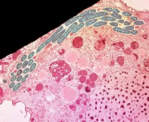
Background imageLegionella Pneumophila Collection: Legionella infected protozoan, TEM C016 / 9412

Legionella infected protozoan, TEM C016 / 9412
Legionella infected protozoan. Transmission electron micrograph (TEM) of a section through a single-celled Tetrahymena pyriformis protozoan, infected with Legionella pneumophila bacteria (yellow)

Background imageLegionella Pneumophila Collection: Hartmannella amoeba and bacterium

Hartmannella amoeba and bacterium. Computer artwork of a Hartmannella vermiformis amoeba (green) engulfing a Legionella pneumophila bacterium (orange) with a pseudopodium

Background imageLegionella Pneumophila Collection: Legionella bacteria, SEM

Legionella bacteria, SEM
Legionella bacteria. Coloured scanning electron micrograph (SEM) of Legionella pneumophila bacteria, the cause of Legionnaires disease

Background imageLegionella Pneumophila Collection: Legionella bacteria, light micrograph

Legionella bacteria, light micrograph
Legionella bacteria. Coloured light micrograph of Legionella pneumophila bacteria, the cause of Legionnaires disease. This rod-shaped

Background imageLegionella Pneumophila Collection: Legionella bacterium, TEM

Legionella bacterium, TEM
Legionella bacteria. Coloured transmission electron micrograph of a Legionella pneumophila bacterium, the cause of Legionnaires disease

Background imageLegionella Pneumophila Collection: Legionella pneumophila bacteria

Legionella pneumophila bacteria

Background imageLegionella Pneumophila Collection: SEM of a colony of Legionella pneumophila

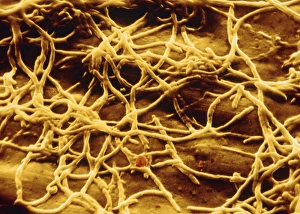
SEM of a colony of Legionella pneumophila
False-colour scanning electron micrograph (SEM) of a colony of Legionella pneumophila, the bacteria causing Legionnaires disease, a severe & often fatal form of lobar pneumonia

Background imageLegionella Pneumophila Collection: SEM of legionella pneumophila

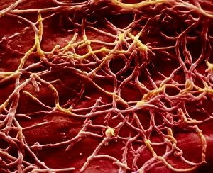
SEM of legionella pneumophila
False-colour scanning electron micrograph (SEM) of a no. of highly-curved forms of the bacterium Legionella pneumophila, the cause of Legionnaires disease, a usually fatal form of lobar pneumonia

Background imageLegionella Pneumophila Collection: Legionella bacteria

Legionella bacteria. Coloured transmission electron micrograph (TEM) of four Legionella pneumophila bacteria. The bacteria (pale green) cause Legionnaires disease


All Professionally Made to Order for Quick Shipping

"Unveiling the Transformation: Legionella pneumophila, from Saul's Conversion to Microscopic Marvels" Witness the remarkable journey of Legionella pneumophila, a bacterium that shares its name with historical events and leaves an indelible mark on scientific exploration. Just as Saint Paul experienced a life-altering conversion, this microbe has captivated researchers worldwide. Delving into the microscopic realm, we encounter stunning views of Legionella pneumophila. Like the Defeat of the Cimbri at Vercellae in 101 B. C. , this bacterium showcases its power under high magnification. Its intricate structure is revealed through multiple microscopic lenses, each unveiling new dimensions. Behold. A captivating sight unfolds as we witness a microscopic view infecting protozoan hosts like Hartmannella amoeba. This symbiotic relationship sheds light on how these bacteria thrive within their chosen habitats. Through scanning electron microscopy (SEM), we uncover mesmerizing images showcasing the elegance and complexity of Legionella bacteria. These snapshots offer glimpses into their world – a world where survival strategies intertwine with beauty. Even under light microscopy, Legionella pneumophila shines brightly as it reveals itself in all its glory. The vibrant colors and distinct patterns captured by these techniques emphasize its significance in both medical research and environmental studies. Finally, our journey concludes with a closer look using transmission electron microscopy (TEM). Here lies an exquisite portrayal of individual cells – every detail meticulously preserved for scrutiny. The intricacies uncovered shed light on how this bacterium operates at such minute scales. Legionella pneumophila may share its name with historical events but make no mistake; it stands tall among scientific marvels today. From ancient tales to modern investigations, this microbe continues to intrigue scientists seeking answers about infectious diseases and microbial ecosystems alike.