
Lateral View Collection
"Exploring the Lateral View: Unveiling Headaches and X-ray Artwork" Delve into the fascinating world of lateral view, where hidden mysteries unfold


All Professionally Made to Order for Quick Shipping
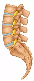
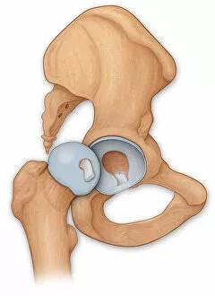
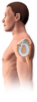
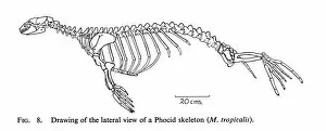
"Exploring the Lateral View: Unveiling Headaches and X-ray Artwork" Delve into the fascinating world of lateral view, where hidden mysteries unfold. From headaches to mesmerizing x-ray artwork, this captivating perspective offers a unique glimpse into various realms. Embark on an intriguing journey as you discover the intricate details of a tonguestone resembling a shark's tooth, adorned with lateral denticles that add an enigmatic touch. Marvel at skeletal structures brought to life through mesmerizing x-ray artwork, revealing the delicate intricacies beneath our skin. Transport yourself back in time with a remarkable 1844 artwork showcasing cervical spinal nerves. Witness how artists from centuries ago meticulously captured anatomical wonders, providing us with invaluable knowledge even today. Step further into history with an enchanting engraving from 1914 depicting "Anatomical View of the Human Torso. " Let your imagination wander as you explore every contour and crevice, appreciating the beauty and complexity of our physical form. Witness normalcy in its purest form through images capturing lumbar vertebrae displaying spinal nerve roots or a man's head and neck accompanied by his skull. These ordinary yet extraordinary views remind us of the marvels within ourselves. Immerse yourself in another dimension as you encounter an adult skull viewed laterally—a testament to human anatomy's intricate design. Observe open hips revealing articular surfaces on femurs—an insight into joint mechanics that keeps us moving effortlessly. Finally, witness arthroscopic surgical repair on a shoulder joint—proof that medical advancements continue to push boundaries in restoring health and mobility for individuals worldwide. Intriguingly diverse yet interconnected, these glimpses into lateral view offer endless fascination for those curious about our bodies' inner workings. Explore this captivating realm where science meets artistry; it is sure to leave you awe-inspired and yearning for more knowledge about our wondrous selves.