
Lamina Propria Collection
The lamina propria is a crucial component of the anatomy and structure of various organs in our body


All Professionally Made to Order for Quick Shipping
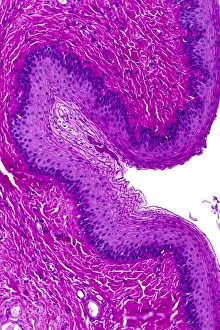
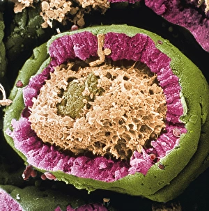
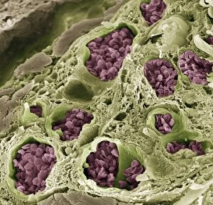
The lamina propria is a crucial component of the anatomy and structure of various organs in our body. Let's explore its significance in different parts, starting with the stomach wall. In the stomach wall, the lamina propria forms one of the layers alongside other structures. It lies beneath the epithelium and above the muscularis mucosae. This layer consists of loose connective tissue that supports and nourishes the overlying cells. Moving on to another area, let's focus on vaginal mucosa. When observed under a light microscope, we can see how this delicate tissue is composed of multiple layers including stratified squamous epithelium. The lamina propria here provides support to these layers while allowing for flexibility during various physiological processes. Similarly, when examining sections of the oesophagus wall under a light microscope, we can observe how important the lamina propria is in maintaining structural integrity. It acts as a foundation for other layers present within this organ. Now shifting our attention to small intestine villi from an ileum section seen through colored SEM imaging, we notice that even at such microscopic levels, there exists a well-defined lamina propria surrounding each villus. This layer plays a vital role in absorbing nutrients from digested food passing through these tiny projections. Lastly, let's not forget about its presence within urethral tissues when viewed under a light microscope. Here again, it serves as an essential supportive layer ensuring proper functioning and maintenance of this vital passage.