Fraud Blocker Skip to main content

Labeled Collection (page 5)

"Exploring the World of Labels: From Art to Science" Delve into the intricate rhythms of life with ECGs showcasing a normal heart rate, beautifully captured in artwork

Background imageLabeled Collection: Salt dissolution, artwork

Salt dissolution, artwork
Salt dissolution. Computer artwork showing how sodium chloride (salt) is dissolved in water. At left is a cubic crystal lattice made up of sodium (orange) and chlorine (green) ions

Background imageLabeled Collection: Platelet activation, artwork

Platelet activation, artwork
actication of PRP gel

Background imageLabeled Collection: Myosin structure, artwork

Myosin structure, artwork
Myosin structure. Computer artwork showing the structure of myosin II, a molecular motor responsible for muscle contraction. Myosin is composed of two heavy chains and four light chains

Background imageLabeled Collection: Bone growth, artwork

Bone growth, artwork
Bone growth. Computer artwork showing the development and growth of a long bone. Behind the epiphysis (head) is an area of spongy bone consisting of trabeculae filled with bone marrow (green)

Background imageLabeled Collection: Muscle anatomy, artwork

Muscle anatomy, artwork
Muscle anatomy. Computer artwork of a cross-section through a skeletal muscle. Single muscle fibres (top right) are made up of many myofibrils surrounded by endomysium connective tissue (orange)

Background imageLabeled Collection: Cephalopd eye, artwork

Cephalopd eye, artwork
Cephalopd eye. Computer artwork showing a cross-section through the eye of a cephalopod. The cephalopod eye is very similar to the vertebrate eye, although the two arose independently

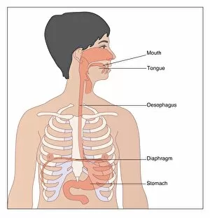
Background imageLabeled Collection: Upper digestive system, artwork

Upper digestive system, artwork
Upper digestive system, computer artwork. At top is the mouth, from which food passes into the oesophagus (gullet), a tube that leads down into the stomach

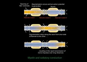
Background imageLabeled Collection: Saltatory conduction of nerve impulse

Saltatory conduction of nerve impulse, computer artwork. Nerve impulses move along myelinated nerves by saltatory conduction. The myelin (orange, striped) does not cover the entire axon

Background imageLabeled Collection: Lung alveolus, artwork

Lung alveolus, artwork
Lung alveolus, computer artwork. Alveoli are the site of gas exchange in the lungs of mammals, where oxygen is taken up and carbon dioxide released

Background imageLabeled Collection: Acrosome reaction, artwork

Acrosome reaction, artwork
Acrosome reaction. Computer artwork showing the process by which an acrosome from a spermatozoon (sperm cell) binds to an ovum (egg cell, bottom). Each spermatozoon has an acrosome in its tip

Background imageLabeled Collection: Heart anatomy, artwork

Heart anatomy, artwork. The heart is a hollow muscle that pumps blood around the body. Deoxygenated blood from the body is carried into the right side of the heart (left) via the vena cava

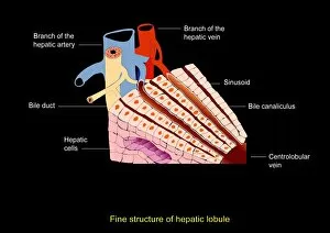
Background imageLabeled Collection: Liver anatomy, artwork

Liver anatomy, artwork
Liver anatomy. Computer artwork of the structure of part of a liver lobule. Oxygenated blood is supplied to the hepatocytes (liver cells) by the hepatic artery (blue)

Background imageLabeled Collection: Eccrine sweat gland, artwork

Eccrine sweat gland, artwork
Eccrine sweat gland, computer artwork. This is one of two types of sweat gland found in the skin. Eccrine glands are found all over the body and release sweat, which consists of water and salt

Background imageLabeled Collection: Echinoderm anatomy, artwork

Echinoderm anatomy, artwork
Echinoderm anatomy. Computer artwork showing the body structure and main organs of a typical echinoderm, such as a starfish

Background imageLabeled Collection: Annelid worm anatomy

Annelid worm anatomy. Computer artwork showing the internal anatomy of 3 segments (metameres) from an annelid (segmented) worms body

Background imageLabeled Collection: DNA nucleotides, artwork

DNA nucleotides, artwork
DNA nucleotides, computer artwork. Section of a double stranded DNA (deoxyribonucleic acid) molecule showing the molecular structure of guanine (G) and cytosine (C) bonded together

Background imageLabeled Collection: Endocrine system, artwork

Endocrine system, artwork
Endocrine system. Computer artwork showing the locations of hormone-producing glands (endocrine glands) in the human body

Background imageLabeled Collection: Insect anatomy, artwork

Insect anatomy, artwork
Insect anatomy. Computer artwork of a cross-section through the body of a typical Insect showing the main structures and organs. For the labelled diagram see image: C008/8724

Background imageLabeled Collection: Exocrine gland types, artwork

Exocrine gland types, artwork
Exocrine gland types. Computer artwork of the six forms an exocrine gland can take. Clockwise from bottom left these are; simple acinar, simple tubular, simple coiled tubular, simple branched acinar

Background imageLabeled Collection: Electrophoresis apparatus, artwork

Electrophoresis apparatus, artwork
Electrophoresis apparatus. Computer artwork showing the apparatus used to separate DNA (deoxyribonucleic acid) fragments using electrophoresis

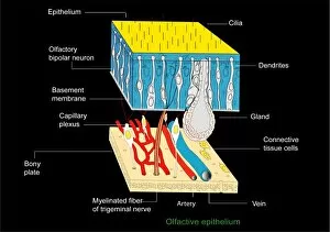
Background imageLabeled Collection: Olfactory epithelium, artwork

Olfactory epithelium, artwork
Olfactory epithelium. Computer artwork showing the structure of the specialised layer of tissue that lines the inside of the nasal cavity and is involved in smell

Background imageLabeled Collection: Ovarian cycle, artwork

Ovarian cycle, artwork
Ovarian cycle, computer artwork. Once sexual maturity is reached up to 20 oocytes start developing into eggs (ovums) every menstrual cycle

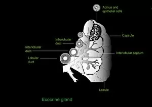
Background imageLabeled Collection: Exocrine gland, artwork

Exocrine gland, artwork
Exocrine gland. Computer artwork of a section through an exocrine gland. Exocrine glands secrete their products through ducts on to epithelial surfaces

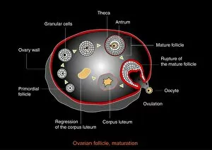
Background imageLabeled Collection: Ovarian follicle maturation, artwork

Ovarian follicle maturation, artwork
Ovarian follicle maturation. Computer artwork showing the beginning of the follicular phase of ovarian follicle maturation. At bottom left is the primary follicle stage

Background imageLabeled Collection: Fraunhofer lines, diagram

Fraunhofer lines, diagram
Fraunhofer lines. Diagram of the Suns spectrum, showing a set of dark absorption lines known as Fraunhofer lines. These are named after the German physicist Joseph von Fraunhofer (1787-1826)

Background imageLabeled Collection: Fruit formation, artwork

Fruit formation, artwork
Fruit formation, computer artwork. At left is a carpel, the female reproductive structure of a plant. A pollen tube (red line) has grown down from a pollen grain (male sex cell)


All Professionally Made to Order for Quick Shipping

"Exploring the World of Labels: From Art to Science" Delve into the intricate rhythms of life with ECGs showcasing a normal heart rate, beautifully captured in artwork. Uncover the wonders of our vision as we unravel the mysteries behind eye anatomy through stunning artistic representations. Embark on an awe-inspiring journey through time and space with a captivating universe timeline depicted in mesmerizing artwork. Witness history being made as you trace the orbit of Sputnik 1, Soviet's groundbreaking achievement in 1957, illustrated in a detailed diagram. Dive into the microscopic world and marvel at Trypanosome protozoan brought to life through exquisite artwork. Discover the building blocks of our body's strength - collagen synthesis and assembly - portrayed artistically for your visual pleasure. Get up close and personal with fibroblast cells, essential contributors to tissue repair and regeneration, showcased in stunning artwork. Experience the beauty behind muscle contraction as it is elegantly depicted through artistry that captures its power and grace. Find solace amidst thorns with Gillray's iconic engraving "Comfort of a Bed of Roses, " offering a satirical take on life's challenges during his era (1756-1815). Explore human anatomy from every angle as you delve into lower back muscles intricately labeled in an illustrative masterpiece. Peer inside animal cell structures like never before, unlocking their secrets through visually striking depictions that captivate your imagination. Relive history with a poignant photograph capturing "The Situation at the Battle of Wounded Knee" when tensions erupted between Native Americans and U. S forces in 1891. Step into this world where art meets science; let these labeled artworks guide you on an extraordinary voyage filled with knowledge, inspiration, and wonderment.