
Ion Channel Collection
"Ion Channels: Unlocking the Secrets of Cellular Communication" Ion channels play a crucial role in various physiological processes


All Professionally Made to Order for Quick Shipping
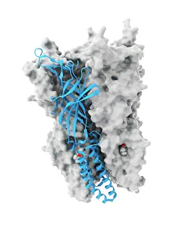
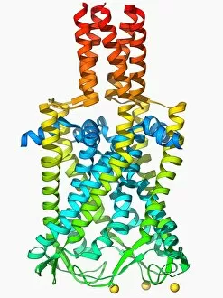
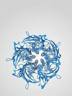
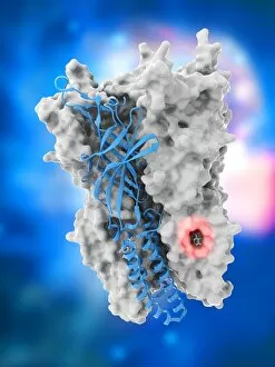
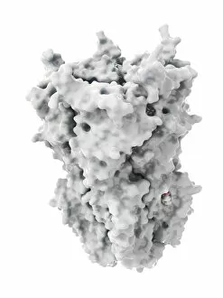
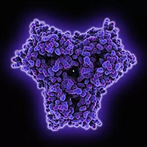
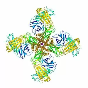
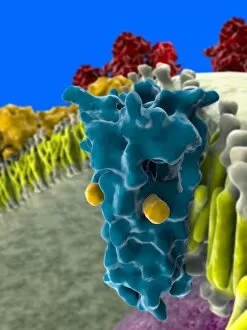
"Ion Channels: Unlocking the Secrets of Cellular Communication" Ion channels play a crucial role in various physiological processes, and their intricate structures hold fascinating secrets. One such example is an anaesthetic inhibiting an ion channel (C015/6718), which sheds light on how these channels can be modulated for medical purposes. The MscL ion channel protein structure reveals its unique architecture, allowing it to respond to mechanical stress and regulate osmotic pressure within cells. On the other hand, the potassium ion channel protein structure showcases its selective permeability to potassium ions, vital for maintaining cellular homeostasis. Delving deeper into potassium ion channels, we discover the importance of the beta subunit in fine-tuning their activity. This subunit acts as a regulatory component that modifies channel kinetics and enhances their functionality. Another intriguing member of this family is the KCNQ ion channel protein structure. Its distinct arrangement enables it to control neuronal excitability and plays a critical role in hearing and epilepsy. Exploring further, we encounter the MscS ion channel protein structure (F006/9650). This mechanosensitive channel responds to membrane tension changes by opening or closing, ensuring cell survival under varying environmental conditions. Intriguingly, studying the cavity structure within potassium ion channels provides insights into how ions navigate through these narrow pathways while maintaining selectivity and efficient transport across cell membranes. Continuing our journey through structural wonders, we revisit MscL's protein structure (F006/9624) alongside MscS's counterpart (F006/9626). These two proteins demonstrate remarkable adaptability in responding to different stimuli while preserving cellular integrity. Lastly, let us not forget about voltage-gated potassium channels (F006/9642), responsible for generating action potentials in excitable cells like neurons and muscle fibers. Their precise conformation allows them to open or close upon changes in membrane potential with exceptional speed and accuracy.