Fraud Blocker Skip to main content

Intestine Collection

"The Intricate World of Intestines: Exploring the Marvels Within" Delving into the depths of our gastrointestinal system

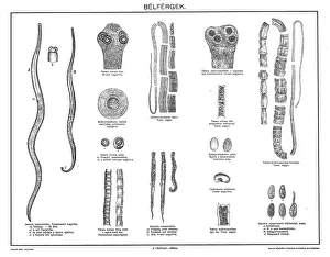
Background imageIntestine Collection: Gastrointestinal nematodes

Gastrointestinal nematodes
Illustration of a gastrointestinal nematodes

Background imageIntestine Collection: Acupuncture

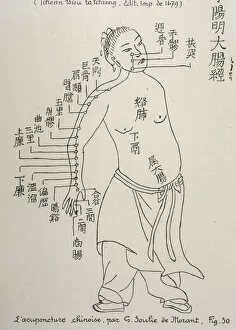
Acupuncture
ACUPUNCTURE The Meridian of the Large Intestine

Background imageIntestine Collection: Deer anatomy, artwork

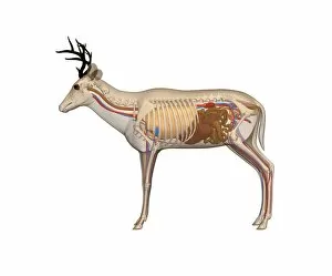
Deer anatomy, artwork
Deer anatomy. Computer artwork showing the internal anatomy of a deer (family Cervidae). Anatomical features shown here include the skeleton, lungs, intestines, bladder

Background imageIntestine Collection: Mosquito (Culicidae), female, internal anatomy, and sucking blood from skin, cross-section

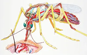
Mosquito (Culicidae), female, internal anatomy, and sucking blood from skin, cross-section

Background imageIntestine Collection: Illustration of greater omentum, or epiploon

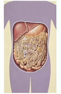
Illustration of greater omentum, or epiploon
Medicine: Human anatomy, greater omentum, or epiploA

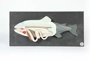
Background imageIntestine Collection: Fish anatomy, historical model

Fish anatomy, historical model
Fish anatomy. 1950s anatomical model showing the internal organs of a typical fish

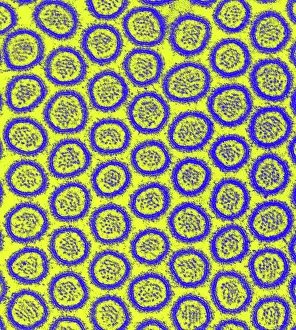
Background imageIntestine Collection: Intestinal microvilli, TEM

Intestinal microvilli, TEM
Intestinal microvilli, coloured transmission electron micrograph (TEM). The microvilli are finger-like projections (seen here in circular cross-section)

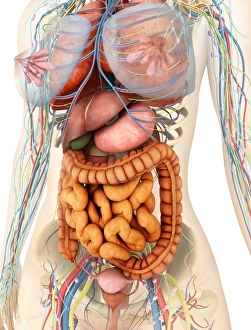
Background imageIntestine Collection: Female body showing digestive and circulatory system

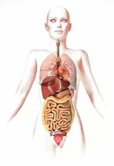
Female body showing digestive and circulatory system
Medical illustration of female body showing digestive and circulatory system, perspective view

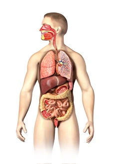
Background imageIntestine Collection: Anatomy of male respiratory system and internal organs

Anatomy of male respiratory system and internal organs
Anatomy of male respiratory system, including the mouth, cutaway view. Other internal organs are visible in half tones

Background imageIntestine Collection: Perspective view of human body, whole organs and bones

Perspective view of human body, whole organs and bones

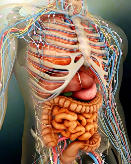
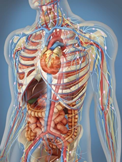
Background imageIntestine Collection: Human body with internal organs, nervous system, lymphatic system and circulatory system

Human body with internal organs, nervous system, lymphatic system and circulatory system
Transparent human body with internal organs, nervous system, lymphatic system and circulatory system

Background imageIntestine Collection: Anatomy of female body with internal organs

Anatomy of female body with internal organs

Background imageIntestine Collection: Internal organs of the respiratory and digestive system

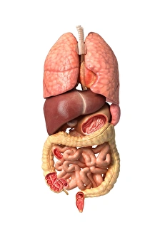
Internal organs of the respiratory and digestive system
Human male anatomy showing internal organs of the respiratory and digestive system

Background imageIntestine Collection: BARTHOLIN, Thomas (1616-1680). Danish physician, mathematician, and theologian

BARTHOLIN, Thomas (1616-1680). Danish physician, mathematician, and theologian
CUL4875726 BARTHOLIN, Thomas (1616-1680). Danish physician, mathematician, and theologian. He is best known for his work in the discovery of the lymphatic system in humans

Background imageIntestine Collection: Midsection view showing internal organs of human body

Midsection view showing internal organs of human body

Background imageIntestine Collection: Human digestive system, with labels

Human digestive system, with labels

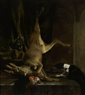
Background imageIntestine Collection: A Dog and a Cat near a partially disembowelled Deer, 1645-1660. Creator: Jan Baptist Weenix

A Dog and a Cat near a partially disembowelled Deer, 1645-1660. Creator: Jan Baptist Weenix
A Dog and a Cat near a partially disembowelled Deer, 1645-1660

Background imageIntestine Collection: The Reward of Cruelty, 1750. Creator: John Bell

The Reward of Cruelty, 1750. Creator: John Bell
The Reward of Cruelty, 1750

Background imageIntestine Collection: Female organs, cut away, artwork

Female organs, cut away, artwork
Female organs, cut away computer artwork

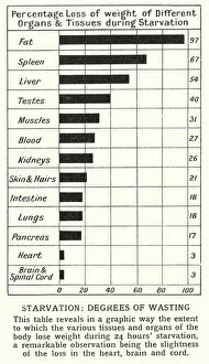
Background imageIntestine Collection: Starvation, degrees of wasting (litho)

Starvation, degrees of wasting (litho)
5998374 Starvation, degrees of wasting (litho) by English School, (20th century); Private Collection; (add.info.: Starvation, degrees of wasting)

Background imageIntestine Collection: Mahasiddha Manibhadra Under a Mango Tree with an Evicerated Dog, 13th / 14th century

Mahasiddha Manibhadra Under a Mango Tree with an Evicerated Dog, 13th / 14th century
Mahasiddha Manibhadra Under a Mango Tree with an Evicerated Dog, 13th/14th century

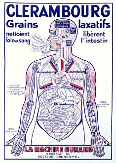
Background imageIntestine Collection: Advertisement for the laxative Clerambourg, early twentieth century

Advertisement for the laxative Clerambourg, early twentieth century
CHT231090 Advertisement for the laxative Clerambourg, early twentieth century (colour litho) by French School, (20th century); Private Collection; (add.info)

Background imageIntestine Collection: MS Hunter 658 Plate XXVI Drawing from William Hunters (1718-83)

MS Hunter 658 Plate XXVI Drawing from William Hunters (1718-83)
GUL232057 MS Hunter 658 Plate XXVI Drawing from William Hunters (1718-83) Anatomy of the Human Gravid Uterus, 1774 (chalk on paper) by Rymsdyk, Jan van (fl.1750-88/89); Glasgow University Library

Background imageIntestine Collection: Model of a disembowelled woman (wax)

Model of a disembowelled woman (wax)
BEN360182 Model of a disembowelled woman (wax) by Susini, Clemente (1754-1814); Museo della Specola, Florence, Italy; (add.info)

Background imageIntestine Collection: The external surface of the intestines infected with cholera

The external surface of the intestines infected with cholera
CHT236338 The external surface of the intestines infected with cholera, from Anatomie Pathologique du Cholera Morbus, by Nikolai Ivanovich Pirogov (1810-81) published in St

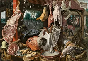
Background imageIntestine Collection: A Meat Stall with the Holy Family Giving Alms, 1551 (oil on panel)

A Meat Stall with the Holy Family Giving Alms, 1551 (oil on panel)
3576758 A Meat Stall with the Holy Family Giving Alms, 1551 (oil on panel) by Aertsen, Pieter (Lange Pier) (1507/08-75); 115.6x168.9 cm; North Carolina Museum of Art, Raleigh

Background imageIntestine Collection: Vintage Anatomical Print of the Human Thoracic and Abdominal Regions, 1912 (screen print)

Vintage Anatomical Print of the Human Thoracic and Abdominal Regions, 1912 (screen print)
3034949 Vintage Anatomical Print of the Human Thoracic and Abdominal Regions, 1912 (screen print) by American School, (20th century); (add.info)

Background imageIntestine Collection: The suicide of the philosopher Cato, who lies on his bed pulling out his innards watched

The suicide of the philosopher Cato, who lies on his bed pulling out his innards watched by horrified disciples. 1648

Background imageIntestine Collection: TYPHUS BACILLUS

TYPHUS BACILLUS
Typhus bacillus, discovered by Eberth, 1880, and studied by Gaffky, 1884 - - left, infected intestine - right, culture

Background imageIntestine Collection: A Butcher, January 1, 1790. January 1, 1790. Creator: Thomas Rowlandson

A Butcher, January 1, 1790. January 1, 1790. Creator: Thomas Rowlandson
A Butcher, January 1, 1790

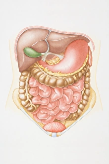
Background imageIntestine Collection: Illustration of abdominal cavity including liver, pancreas, stomach, small intestine

Illustration of abdominal cavity including liver, pancreas, stomach, small intestine, colon and bladder

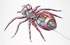
Background imageIntestine Collection: Spider (Araneae), internal anatomy, cross-section

Spider (Araneae), internal anatomy, cross-section

Background imageIntestine Collection: ALASKA: ESKIMO WOMAN. An Eskimo woman wearing a nose ring and and a labret, dressed

ALASKA: ESKIMO WOMAN. An Eskimo woman wearing a nose ring and and a labret, dressed in a hooded parka made of intestinal parchment, Nunivak Island, Alaska. Photographed by Edward S. Curtis, c1929

Background imageIntestine Collection: Anatomy of human digestive system, male representation

Anatomy of human digestive system, male representation

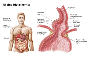
Background imageIntestine Collection: Medical ilustration of a hiatal hernia in the upper part of the stomach into the thorax

Medical ilustration of a hiatal hernia in the upper part of the stomach into the thorax

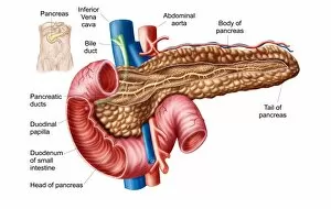
Background imageIntestine Collection: Anatomy of pancreas

Anatomy of pancreas

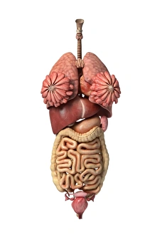
Background imageIntestine Collection: 3D rendering of healthy female internal organs

3D rendering of healthy female internal organs

Background imageIntestine Collection: Anatomy of human abdominal vein system

Anatomy of human abdominal vein system

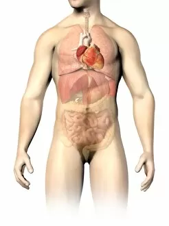
Background imageIntestine Collection: Male anatomy of internal organs

Male anatomy of internal organs, with heart highlighted and rest of organs in half tone

Background imageIntestine Collection: Anatomy of human digestive system, front view

Anatomy of human digestive system, front view

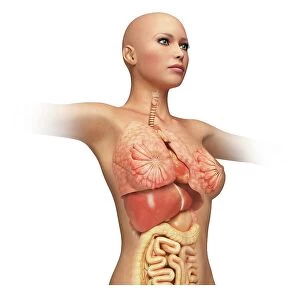
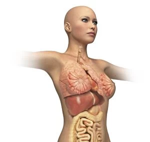
Background imageIntestine Collection: Woman body midsection with interior organs superimposed

Woman body midsection with interior organs superimposed

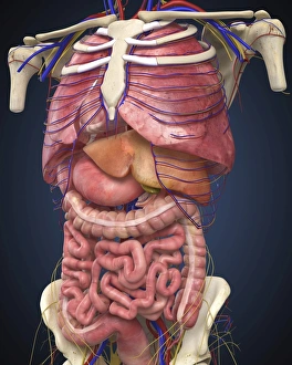
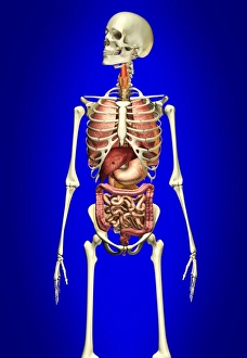
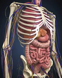
Background imageIntestine Collection: Male skeleton with internal organs on blue background

Male skeleton with internal organs on blue background

Background imageIntestine Collection: Sectional view of large intestine

Sectional view of large intestine

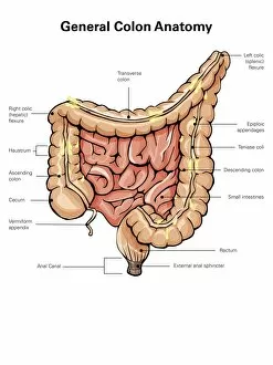
Background imageIntestine Collection: General colon anatomy, with labels

General colon anatomy, with labels

Background imageIntestine Collection: Human midsection with internal organs

Human midsection with internal organs

Background imageIntestine Collection: Cross section of human digestive system

Cross section of human digestive system

Background imageIntestine Collection: Anatomy of female body with internal organs superimposed

Anatomy of female body with internal organs superimposed

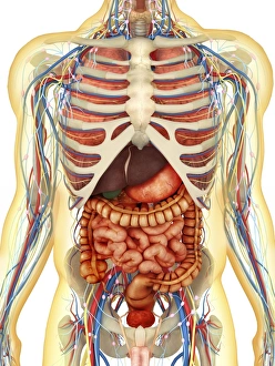
Background imageIntestine Collection: Human body showing heart and main circulatory system position

Human body showing heart and main circulatory system position
Transparent human body showing heart and main circulatory system position with internal organs, nervous system, lymphatic system and circulatory system

Background imageIntestine Collection: Female anatomy, artwork F007 / 3566

Female anatomy, artwork F007 / 3566
Female anatomy, computer artwork

Background imageIntestine Collection: Intestinal villi, SEM

Intestinal villi, SEM
Intestinal villi. Coloured scanning electron micrograph (SEM) of a section through a part of the small intestine, showing the villi that cover its inner surface

Background imageIntestine Collection: Human organs, artwork

Human organs, artwork
Human organs, computer artwork

Background imageIntestine Collection: Capillaria philippinensis, parasitic worm

Capillaria philippinensis, parasitic worm
This worm species (Capillaria philippinensis) is a parasite of the human small intestine

Background imageIntestine Collection: Cow anatomy, artwork

Cow anatomy, artwork
Cow anatomy. Computer artwork showing the internal anatomy of a domesticated cow (Bos primigenius). Anatomical features shown here include the skeleton, lungs, intestines, bladder

Background imageIntestine Collection: Lazio Roma Rome Velletri Museo Civico di Velletri

Lazio Roma Rome Velletri Museo Civico di Velletri
Lazio Roma Velletri Museo Civico di Velletri, this is my Italy, the italian country of visual history, Medieval Marble tondo of the depositon, early christian sarcophagus

Background imageIntestine Collection: Abstract Of Intestines

Abstract Of Intestines

Background imageIntestine Collection: Stomach digestive system engraving 1895

Stomach digestive system engraving 1895
Meyers Konversations-Lexikon. Ein Nachschlagewerk des allgemeinen Wissens, 5th edition 17 volumes Bibliographisches Institut - Leipzig 1895-1897

Background imageIntestine Collection: Illustration of human digestive and respiratory systems

Illustration of human digestive and respiratory systems

Background imageIntestine Collection: Anatomy of human pancreas, with labels

Anatomy of human pancreas, with labels

Background imageIntestine Collection: Conceptual image of internal organs of the respiratory and digestive systems

Conceptual image of internal organs of the respiratory and digestive systems

Background imageIntestine Collection: Comparison of healthy and unhealthy female organs

Comparison of healthy and unhealthy female organs
3D rendering comparing healthy female organs and unhealthy female organs

Background imageIntestine Collection: 3D rendering of unhealthy female internal organs

3D rendering of unhealthy female internal organs

Background imageIntestine Collection: Human body and digestive system, perspective view

Human body and digestive system, perspective view

Background imageIntestine Collection: Medical illustration of fetus development at 36 weeks

Medical illustration of fetus development at 36 weeks

Background imageIntestine Collection: Female anatomy, artwork F007 / 1849

Female anatomy, artwork F007 / 1849
Female anatomy, computer artwork

Background imageIntestine Collection: Female digestive system, artwork

Female digestive system, artwork
Female digestive system, computer artwork

Background imageIntestine Collection: Pancreatic acinar cell

Pancreatic acinar cell. Transmission electron micrograph (TEM) of a section through an enzyme-secreting acinar cell in the human pancreas, showing part of the nucleus (round, far left)

Background imageIntestine Collection: Goblet cell, TEM

Goblet cell, TEM
Goblet cell. Transmission electron micrograph (TEM) of a section through a goblet cell in the lining (epithelium, top) of the small intestine

Background imageIntestine Collection: Gastric antral vascular ectasia C016 / 8328

Gastric antral vascular ectasia C016 / 8328
Gastric antral vascular ectasia. Endoscopic view of the antrum (final portion of stomach), affected by gastric antral vascular ectasia (GAVE)

Background imageIntestine Collection: Small intestine, endoscopic view C016 / 8323

Small intestine, endoscopic view C016 / 8323
Small intestine. Endoscopic view of the healthy interior of a persons duodenum - the first part of the small intestine, showing the muscular wall

Background imageIntestine Collection: Colon cancer, endoscopic view C016 / 8308

Colon cancer, endoscopic view C016 / 8308
Colon cancer. Endoscopic view of a malignant (cancerous) adenocarcinoma (upper right) in a patients ascending colon (large intestine)

Background imageIntestine Collection: ANATOMY: TORSO. General view of the organs of the thorax and abdomen. Engraving, 19th century

ANATOMY: TORSO. General view of the organs of the thorax and abdomen. Engraving, 19th century

Background imageIntestine Collection: Earthworm, transverse section

Earthworm, transverse section
Earthworm. Light micrograph of a transverse section through the body of a round segmented earthworm (Lumbricus terrestris) in the intestinal region

Background imageIntestine Collection: Healthy small intestines, artwork

Healthy small intestines, artwork
Healthy small intestines, computer artwork

Background imageIntestine Collection: The Harmful Effects of the Corset (Phenomenes chimiques de la respiration)

The Harmful Effects of the Corset (Phenomenes chimiques de la respiration)
The Harmful Effects of the Corset (Phe nome nes chimiques de la respiration), illustration from La Vie Normale et la Sante by Dr Jules Rengade, pub. 1881 (coloured engraving)

Background imageIntestine Collection: Diagram showing the Alimentary Canal, which passes right through the body, c1934

Diagram showing the Alimentary Canal, which passes right through the body, c1934. From The Treasury of Knowledge. [Richard Clay & Sons, Ltd. Bungay, Suffolk, 1934]

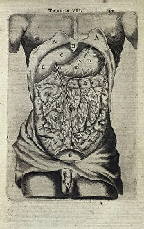
Background imageIntestine Collection: Abdominal cavity and its contents, 1543

Abdominal cavity and its contents, 1543. From De humani corporis fabrica by Andreas Vesalius. (Basle, 1543)

Background imageIntestine Collection: VALUE

VALUE
Human Anatomy. Digestive system. Engraving


All Professionally Made to Order for Quick Shipping

"The Intricate World of Intestines: Exploring the Marvels Within" Delving into the depths of our gastrointestinal system, we uncover a world teeming with life - intestinal parasites known as gastrointestinal nematodes. Did you know that acupuncture can have a positive impact on our intestines? This ancient practice stimulates specific points to promote digestive health and balance. A female mosquito (Culicidae) is not just an annoying pest; its internal anatomy reveals fascinating adaptations for sucking blood from our skin, as seen in a cross-section illustration. The greater omentum, or epiploon, acts as a protective apron within our abdomen. An intriguing historical model showcases its intricate folds and functions. Intestinal protozoan parasites are microscopic organisms that wreak havoc on our gut health. Transmission electron microscopy (TEM) captures their detailed structure and complexity. Parasitic nematode worms may sound like creatures from a horror movie, but they inhabit the intestines of various animals including humans. Understanding their lifecycle is crucial for prevention and treatment. Cryptosporidium protozoa are notorious culprits behind diarrheal diseases worldwide. TEM images provide insights into their unique features and strategies for survival within the intestine. Tiny but mighty - intestinal microvilli play a vital role in nutrient absorption by increasing surface area in the small intestine's lining, as revealed through TEM imaging. Explore an artistic representation of dog anatomy where the intricacies of their intestines come to life through stunning artwork capturing both form and function. The Lazio Roma Rome Velletri Museo Civico di Velletri houses captivating exhibits showcasing ancient Roman medical knowledge related to intestinal health – truly bridging past with present discoveries.

© Copyright Media Storehouse, All Rights Reserved 2003 - 2024
Facebook     Pinterest     Card Payments     Reviews IO