
Insulin Collection
Insulin, a vital hormone produced by the pancreas, plays a crucial role in regulating blood sugar levels


All Professionally Made to Order for Quick Shipping
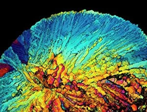
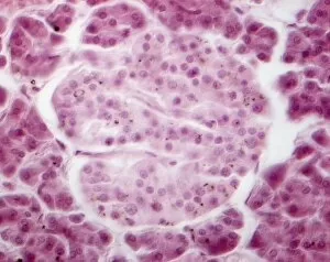
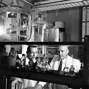
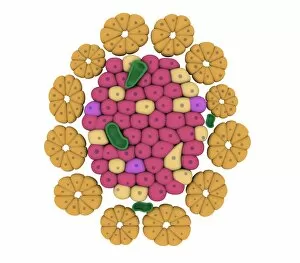
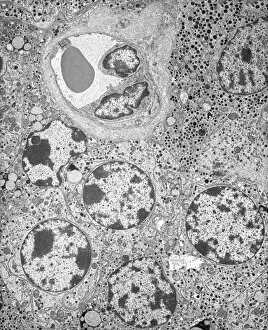
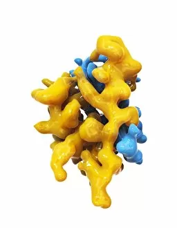
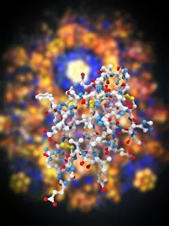
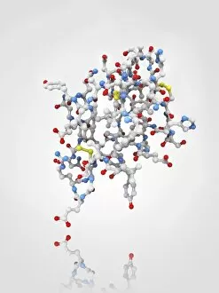
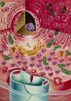
Insulin, a vital hormone produced by the pancreas, plays a crucial role in regulating blood sugar levels. The intricate anatomy of the pancreas houses the remarkable Islets of Langerhans, where insulin is synthesized and secreted. These microscopic clusters of cells are beautifully captured in light micrographs, showcasing their significance. The discovery was a monumental breakthrough in medical history. Canadian physiologists Charles Herbert Best and Frederick Grant Banting were instrumental in unraveling its secrets. In 1923, Banting and his team successfully isolated insulin from pancreatic extracts, revolutionizing diabetes treatment forever. Sir Frederick Grant Banting's contributions to medicine extend beyond his co-discovery of insulin; he was also an accomplished painter and Nobel laureate. Gordon Ross' illustration vividly portrays this multi-talented scientist who left an indelible mark on humanity through his groundbreaking work. Artwork depicting insulin crystals further emphasizes its importance as a therapeutic agent for individuals with diabetes. Insulin injections have become a lifeline for millions worldwide, enabling them to manage their condition effectively. These artistic renderings capture the essence of this life-saving procedure. A cross-sectional biomedical illustration sheds light on how sulphonylurea drugs stimulate insulin-producing cells within the pancreas to increase production. This innovative approach has provided new avenues for managing diabetes and improving patients' quality of life. In summary, from its intricate anatomy within the pancreas to its crystalline structure under microscopic examination, insulin remains at the forefront of medical advancements thanks to brilliant minds like Best and Banting. As we continue our quest for better treatments for diabetes, let us appreciate both the scientific marvels behind this hormone and those who dedicated their lives to understanding it fully.