Fraud Blocker Skip to main content

Infected Collection (page 20)

"Unveiling the Hidden World of Infection

Background imageInfected Collection: Chlamydia, SEM

Chlamydia, SEM
Chlamydia. Coloured scanning electron micrograph (SEM) of chlamydia (Chlamydia sp.) bacteria (green, spherical, centre) in a ruptured cell

Background imageInfected Collection: Chlamydia bacteria, SEM

Chlamydia bacteria, SEM
Chlamydia bacteria. Coloured scanning electron micrograph of Chlamydia trachomatis bacteria

Background imageInfected Collection: Bacterial contamination, SEM

Bacterial contamination, SEM
Bacterial contamination, coloured scanning electron micrograph (SEM). Escherichia coli bacteria in a cell culture. This contamination has come from an unclean water source

Background imageInfected Collection: Infected nail, SEM

Infected nail, SEM
Bacteria on a nail. Coloured scanning electron micrograph (SEM) of a nail with a bacterial infection (paronychia). Treatment is with antibiotic drugs

Background imageInfected Collection: Epithelial gonorrhoea infection, SEM

Epithelial gonorrhoea infection, SEM
Epithelial gonorrhoea infection. Coloured scanning electron micrograph (SEM) of Neisseria gonorrhoeae bacteria (clusters of small round cells) and an epithelial cell (large structure, centre)

Background imageInfected Collection: E. coli and Shigella sp. bacteria

E. coli and Shigella sp. bacteria. Fluorescence confocal light micrograph of Escherichia coli and Shigella sp. bacteria (blue) in human Caco-2 cells (green)

Background imageInfected Collection: Fungus on maple leaf

Fungus on maple leaf. Unidentified fungus (red) growing on the tips of maple (Acer sp.) leaves. Photographed in the Lower Stein Valley, southern British Columbia, Canada

Background imageInfected Collection: Tar spot fungus on sycamore leaves

Tar spot fungus on sycamore leaves. Leaves of a sycamore (Acer pseudoplatanus) tree, affected by the fungal pathogen Rhytisma acerinum, which causes a disease known as tar spot

Background imageInfected Collection: Cell infected with HIV, SEM

Cell infected with HIV, SEM
Cell infected with HIV. Coloured scanning electron micrograph (SEM) of HIV particles (yellow) budding from the membrane of a host cell (red)

Background imageInfected Collection: Athletes foot fungus, SEM

Athletes foot fungus, SEM
Athletes foot. Coloured scanning electron micrograph (SEM) of spores (yellow) of the fungus that causes athletes foot (tinea pedis) on skin (purple) from a human foot

Background imageInfected Collection: Palm seeds infected with pestalotia

Palm seeds infected with pestalotia
Palm seeds infected with the parasitic fungus Pestalotia sp. (black growths)

Background imageInfected Collection: Diphtheria at Woolwich

Diphtheria at Woolwich
Sketches of the diphtheria huts on Woolwich Common. The huts were used to confine those infected by the epidemic

Background imageInfected Collection: St Roch as a Pilgrim

St Roch as a Pilgrim
(Rocco, Roque) - He goes as a pilgrim to Rome, where he is infected while caring for plague victims; he recovers and goes on to perform many healing miracles

Background imageInfected Collection: Roman Cattle Stricken

Roman Cattle Stricken
Cattle farming : the herd struck by cattle plague - slaughtering the infected animals

Background imageInfected Collection: Illegal weapon seized by Cumbria Police UK

Illegal weapon seized by Cumbria Police UK
Ashley Cooper / SpecialistStock

Background imageInfected Collection: Rabbit in late stages of Myxomatosis showing swelling around eyes, Cotswolds, UK

Rabbit in late stages of Myxomatosis showing swelling around eyes, Cotswolds, UK
MAB-89 Rabbit in late stages of Myxomatosis showing swelling around eyes Cotswolds, UK Mark Boulton Please note that prints are for personal display purposes only

Background imageInfected Collection: Disinfecting a home - 2

Disinfecting a home - 2
DISINFECTING A HOME Infected bedlinen is fumigated in a horse-drawn portable Stove to destroy germs of tuberculosis or cholera

Background imageInfected Collection: Bubonic Plague, Portugal

Bubonic Plague, Portugal
When Portugal is struck by an epidemic of bubonic plague, the population burn infected homes in hope of destroying the source of the trouble


All Professionally Made to Order for Quick Shipping

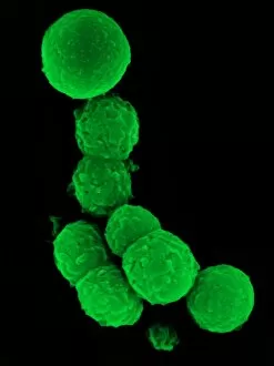
"Unveiling the Hidden World of Infection: A Glimpse into Medical History and Microscopic Marvels" Step back in time to the 19th century as we explore the fascinating world of infection. Calots spinal surgery, a groundbreaking procedure for its time, left surgeons grappling with the challenges of preventing post-operative infections. In another breakthrough, tuberculosis detection took a leap forward with the advent of X-ray technology. This revolutionary method allowed doctors to visualize the disease within patients' lungs, aiding in diagnosis and treatment. Delving deeper into microscopic realms, we encounter head lice through scanning electron microscopy (SEM). These tiny parasites that plagued humanity throughout history come to life under high magnification. Moving on from lice to bacteria, SEM reveals E. Coli's intricate structure. The detailed images showcase these notorious pathogens responsible for various illnesses and foodborne outbreaks. Shifting our focus to bacterial meningitis, magnetic resonance imaging (MRI) scans provide invaluable insights into this dangerous infection affecting the brain and spinal cord. The non-invasive nature of MRI aids in early detection and prompt intervention. Venturing outdoors brings us face-to-face with sandflies—a carrier of diseases such as leishmaniasis—captured through powerful microscopes that expose their delicate features. Witnessing phagocytosis at work is truly awe-inspiring when observing fungal spores being engulfed by immune cells under SEM. This process showcases our body's defense mechanism against harmful invaders. Transitioning from fungi to viruses, transmission electron microscopy (TEM) unravels hepatitis C viruses lurking within liver cells—an essential tool for understanding viral infections and developing effective treatments. Further exploring TEM's capabilities reveals intestinal protozoan parasites wreaking havoc inside human intestines. These microscopic organisms cause severe gastrointestinal distress but can now be studied more closely than ever before.