Fraud Blocker Skip to main content

Immunological Collection

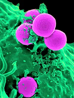
"Unlocking the Power of Immunological Defense: A Visual Journey" Witness the incredible battle between a Neutrophil and MRSA as it engulfs the harmful bacteria

Background imageImmunological Collection: Neutrophil engulfing MRSA, SEM C018 / 8596

Neutrophil engulfing MRSA, SEM C018 / 8596
Neutrophil engulfing MRSA. Coloured scanning electron micrograph (SEM) of a neutrophil white blood cell (green) engulfing methicillin-resistant Staphylococcus aureus bacteria (MRSA, pink)
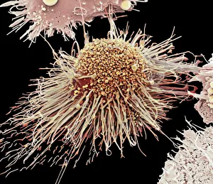
Background imageImmunological Collection: Activated macrophage, SEM C015 / 6375

Activated macrophage, SEM C015 / 6375
Activated macrophage. Coloured scanning electron micrograph (SEM) of an active macrophage white blood cell. Macrophages are cells of the bodys immune system that are found in the tissues rather than

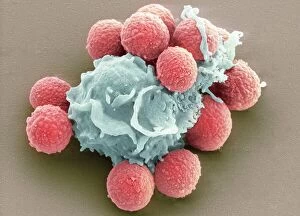
Background imageImmunological Collection: Phagocytosis of fungal spores, SEM

Phagocytosis of fungal spores, SEM
Phagocytosis of a fungus spore. Coloured scanning electron micrograph (SEM) of a neutrophil (blue) phagocytosing (engulfing and destroying) spores from the fungus Aspergillus fumigatus (red)

Background imageImmunological Collection: Lymphocyte white blood cells, artwork

Lymphocyte white blood cells, artwork
Lymphocyte white blood cells. Computer artwork of lymphocyte white blood cells, showing their nuclei (red, centre). Lymphocytes, like all white blood cells, are part of the immune system

Background imageImmunological Collection: White and red blood cells, illustration

White and red blood cells, illustration
3d illustration of white blood cells (leukocytes) in the human body

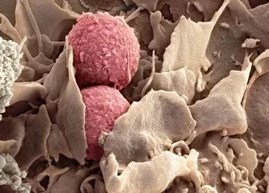
Background imageImmunological Collection: Phagocytosis of fungus spores, SEM

Phagocytosis of fungus spores, SEM
Phagocytosis of fungus spores. Coloured scanning electron micrograph (SEM) of spores from the fungus Aspergillus fumigatus (red) being phagocytosed (engulfed and destroyed) by a dendritic cell (brown)

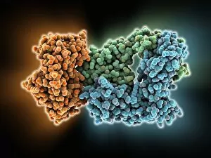
Background imageImmunological Collection: Immunoglobulin G antibody and egg white F006 / 9682

Immunoglobulin G antibody and egg white F006 / 9682
Immunoglobulin G and egg white. Molecular model of an immunoglobulin G (IgG) antibody bound to a molecule of egg white. This is the most abundant immunoglobulin and is found in all body fluids

Background imageImmunological Collection: Thymus gland, light micrograph C015 / 4970

Thymus gland, light micrograph C015 / 4970
Thymus gland. Light micrograph of a section through tissue from the thymus gland, part of the lymphoid system. The dense areas are the thymic cortex, which produces lots of lymphocytes

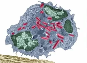
Background imageImmunological Collection: Macrophage and lymphocytes, TEM

Macrophage and lymphocytes, TEM
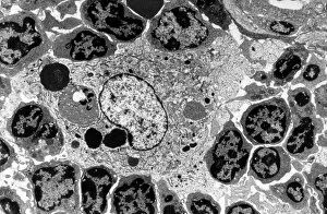
Macrophage and lymphocytes, transmission electron micrograph (TEM). Macrophage surrounded by lymphocytes in a lymph node. This view includes many intercellular contacts

Background imageImmunological Collection: Immunoglobulin A, molecular model

Immunoglobulin A, molecular model
Immunoglobulin A. Molecular model of the structure of the antibody immunoglobulin A (IgA). This is the secretory dimeric form (sIgA), the main immunoglobulin found in secretions such as saliva

Background imageImmunological Collection: White blood cells and platelets, SEM C016 / 3099

White blood cells and platelets, SEM C016 / 3099
White blood cells and platelets. Coloured scanning electron micrograph (SEM) of platelets (green) with a variety of different white blood cells (leucocytes, yellow)

Background imageImmunological Collection: White blood cells and platelets, SEM C016 / 3098

White blood cells and platelets, SEM C016 / 3098
White blood cells and platelets. Coloured scanning electron micrograph (SEM) of platelets (purple) with a variety of different white blood cells (leucocytes, blue)

Background imageImmunological Collection: Monocyte white blood cell, SEM C016 / 3089

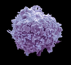
Monocyte white blood cell, SEM C016 / 3089
Monocyte white blood cell. Scanning electron micrograph (SEM) of a monocyte white blood cell. Monocytes, like all white blood cells, are part of the human bodys immune system

Background imageImmunological Collection: Peter Medawar, British immunologist C017 / 7119

Peter Medawar, British immunologist C017 / 7119
Peter Medawar (1915-1987), British immunologist. Medawar was professor of zoology at the University of Birmingham and then University College, London

Background imageImmunological Collection: MRSA and dead neutrophil, SEM C018 / 8601

MRSA and dead neutrophil, SEM C018 / 8601
MRSA and dead neutrophil. Coloured scanning electron micrograph (SEM) of methicillin-resistant Staphylococcus aureus bacteria (MRSA, yellow) and a dead neutrophil white blood cell (red)

Background imageImmunological Collection: Neutrophil engulfing MRSA, SEM C018 / 8597

Neutrophil engulfing MRSA, SEM C018 / 8597
Neutrophil engulfing MRSA. Coloured scanning electron micrograph (SEM) of a neutrophil white blood cell (bottom) engulfing methicillin-resistant Staphylococcus aureus bacteria (MRSA, yellow)

Background imageImmunological Collection: Eosinophil white blood cell, TEM C014 / 1438

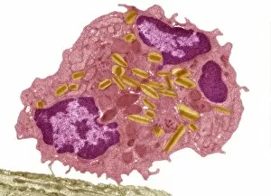
Eosinophil white blood cell, TEM C014 / 1438
Eosinophil white blood cell. Transmission electron micrograph (TEM) of a section through an eosinophil. Eosinophils, like all white blood cells, are part of the bodys immune system

Background imageImmunological Collection: Human antitumour antibody molecule

Human antitumour antibody molecule
Human antitumour antibody. Molecular model showing the antitumour antibody BR96 complexed with part of the Lewis antigen. The Lewis antigen is expressed on the surface of human carcinoma cells

Background imageImmunological Collection: Edward Jenner, British physician C017 / 7108

Edward Jenner, British physician C017 / 7108
Edward Jenner (1749-1823), British physician. Jenner, who also did work as a naturalist, is famed for developing a vaccine for the often fatal viral infection smallpox (syringe in hand)

Background imageImmunological Collection: Tofacitinib rheumatoid arthritis drug F007 / 0196

Tofacitinib rheumatoid arthritis drug F007 / 0196
Tofacitinib rheumatoid arthritis drug, molecular model. Atoms are represented as spheres and are colour-coded: hydrogen (white), carbon (grey), oxygen (red) and nitrogen (blue)

Background imageImmunological Collection: Tofacitinib rheumatoid arthritis drug F007 / 0195

Tofacitinib rheumatoid arthritis drug F007 / 0195
Tofacitinib rheumatoid arthritis drug, molecular model. Atoms are represented as spheres and are colour-coded: hydrogen (white), carbon (grey), oxygen (red) and nitrogen (blue)

Background imageImmunological Collection: White blood cell and platelet, SEM F006 / 9929

White blood cell and platelet, SEM F006 / 9929
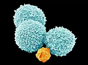
White blood cells. Coloured scanning electron micrograph (SEM) of white blood cells (leucocytes) and a platelet (thrombocyte, red). Magnification: x6, 600 when printed at 10 centimetres wide

Background imageImmunological Collection: White blood cell and platelet, SEM F006 / 9928

White blood cell and platelet, SEM F006 / 9928
White blood cells. Coloured scanning electron micrograph (SEM) of white blood cells (leucocytes) and a platelet (thrombocyte, orange). Magnification: x6, 600 when printed at 10 centimetres wide

Background imageImmunological Collection: White blood cell and platelet, SEM F006 / 9927

White blood cell and platelet, SEM F006 / 9927
White blood cells. Coloured scanning electron micrograph (SEM) of white blood cells (leucocytes) and a platelet (thrombocyte, green). Magnification: x6, 600 when printed at 10 centimetres wide

Background imageImmunological Collection: White blood cells, SEM F006 / 9924

White blood cells, SEM F006 / 9924
White blood cells. Coloured scanning electron micrograph (SEM) of white blood cells (leucocytes). Magnification: x2, 400 when printed at 10 centimetres wide

Background imageImmunological Collection: White blood cells, SEM F006 / 9923

White blood cells, SEM F006 / 9923
White blood cells. Coloured scanning electron micrograph (SEM) of white blood cells (leucocytes). Magnification: x2, 400 when printed at 10 centimetres wide

Background imageImmunological Collection: White blood cells, SEM F006 / 9926

White blood cells, SEM F006 / 9926
White blood cells. Coloured scanning electron micrograph (SEM) of white blood cells (leucocytes). Magnification: x2, 400 when printed at 10 centimetres wide

Background imageImmunological Collection: White blood cells, SEM F006 / 9925

White blood cells, SEM F006 / 9925
White blood cells. Coloured scanning electron micrograph (SEM) of white blood cells (leucocytes). Magnification: x2, 400 when printed at 10 centimetres wide

Background imageImmunological Collection: White blood cells, SEM F006 / 9922

White blood cells, SEM F006 / 9922
White blood cells. Coloured scanning electron micrograph (SEM) of white blood cells (leucocytes). Magnification: x2, 400 when printed at 10 centimetres wide

Background imageImmunological Collection: Eosinophil white blood cell, TEM C014 / 1437

Eosinophil white blood cell, TEM C014 / 1437
Eosinophil white blood cell. Transmission electron micrograph (TEM) of a section through an eosinophil. Eosinophils, like all white blood cells, are part of the bodys immune system

Background imageImmunological Collection: Antibodies and their antigen F006 / 9678

Antibodies and their antigen F006 / 9678
Antibodies and their antigen. Molecular model showing the molecular structure of two anti-P-glycoprotein antibodies (upper left and right) and the P-glycoprotein (v-shaped) to which they respond

Background imageImmunological Collection: Interferon antagonism by viral protein F006 / 9662

Interferon antagonism by viral protein F006 / 9662
Interferon (IFN) antagonism by viral protein. Molecular model of an orthopoxvirus IFN-gamma-binding protein bound to an IFN-gamma molecule

Background imageImmunological Collection: T cell receptor, molecular model F006 / 9515

T cell receptor, molecular model F006 / 9515
T cell receptor. Molecular model of an alpha T cell receptor. T cell receptors are protein complexes found on the surface of a type of white blood cell called T lymphocytes (or T cells)

Background imageImmunological Collection: Flu virus surface protein and antibody F006 / 9428

Flu virus surface protein and antibody F006 / 9428
Flu virus surface protein and antibody. Molecular model of the neuraminidase glycoprotein enzyme found on the surface of the influenza (flu) virus

Background imageImmunological Collection: Immunoglobulin G antibody molecule F006 / 9371

Immunoglobulin G antibody molecule F006 / 9371
Immunoglobulin G (IgG) antibody, molecular model. This is the most abundant immunoglobulin and is found in all body fluids

Background imageImmunological Collection: Human interferon alpha molecule F006 / 9373

Human interferon alpha molecule F006 / 9373
Human interferon alpha, molecular model. Interferons are proteins produced by white blood cells as part of the immune response to invading pathogens, especially viruses

Background imageImmunological Collection: MHC protein-antigen complex F006 / 9363

MHC protein-antigen complex F006 / 9363
MHC protein-antigen complex. Molecular model of the human class I MHC (major histocompatibility complex) protein HLA-B27 complexed with beta-2 microglobulin

Background imageImmunological Collection: Eosinophil white blood cell, TEM C014 / 1439

Eosinophil white blood cell, TEM C014 / 1439
Eosinophil white blood cell. Transmission electron micrograph (TEM) of a section through an eosinophil. Eosinophils, like all white blood cells, are part of the bodys immune system

Background imageImmunological Collection: T cell receptor antigen complex F006 / 9339

T cell receptor antigen complex F006 / 9339
T cell receptor antigen complex. Molecular model of the alphabeta T cell receptor bound to the influenza haemagglutinin antigen and MHC class II molecule HLA-DR1

Background imageImmunological Collection: Birch pollen allergen F006 / 9335

Birch pollen allergen F006 / 9335
Birch pollen allergen. Molecular model of Bet v 1l, the molecule responsible for allergic reactions to birch pollen. Here

Background imageImmunological Collection: MHC protein complexed with flu virus F006 / 9294

MHC protein complexed with flu virus F006 / 9294
MHC protein complexed with flu virus. Molecular model showing human class II MHC (major histocompatibility complex) protein HLA-DR1 complexed with an influenza (flu) virus peptide

Background imageImmunological Collection: CD4 protein molecule F006 / 9266

CD4 protein molecule F006 / 9266
CD4 protein. Molecular model of the CD4 T cell (T lymphocyte) receptor protein. CD4 binds to the surface proteins on other cells and pathogenic micro-organisms

Background imageImmunological Collection: T cell receptor B7 molecule F006 / 9247

T cell receptor B7 molecule F006 / 9247
T cell receptor B7. Molecular model of the T cell receptor B7 bound to the viral Tax peptide and MHC class I molecule HLA-A2

Background imageImmunological Collection: Human interferon beta molecule F006 / 9241

Human interferon beta molecule F006 / 9241
Human interferon beta, molecular model. Interferons are proteins produced by white blood cells as part of the immune response to invading pathogens, especially viruses

Background imageImmunological Collection: Interleukin-6, molecular model F006 / 9232

Interleukin-6, molecular model F006 / 9232
Interleukin-6. Molecular model of the cytokine protein human interleukin-6. This protein is produced in the body and has a wide variety of functions in the immune system

Background imageImmunological Collection: Antibodies and viruses, artwork

Antibodies and viruses, artwork
Antibodies and viruses. Computer artwork showing antibody, or immunoglobulin, molecules (blue) surrounding virus particles (virions, yellow)

Background imageImmunological Collection: T-cell receptor bound to enterotoxin

T-cell receptor bound to enterotoxin, molecular model. The T cell receptor (TCR) is a protein complex found on the surface of a type of white blood cell called T lymphocytes (or T cells)

Background imageImmunological Collection: NK cells attacking cancer cells, artwork

NK cells attacking cancer cells, artwork
NK cells attacking cancer cells. Computer artwork of human Natural Killer (NK) cells (small, round) attacking cancer cells (large, round)


All Professionally Made to Order for Quick Shipping

"Unlocking the Power of Immunological Defense: A Visual Journey" Witness the incredible battle between a Neutrophil and MRSA as it engulfs the harmful bacteria, captured in stunning detail through SEM C018 / 8596. Delve into the intricate world of phagocytosis as fungal spores are devoured by specialized cells, showcased beautifully in an SEM image. Behold the might of an Activated Macrophage, ready to defend our bodies against invading pathogens, magnified through SEM C015 / 6375. Explore the artistic representation of Lymphocyte white blood cells, showcasing their crucial role in immune response with captivating artwork. Marvel at Dendritic cells depicted in breathtaking artwork - these sentinel-like cells play a pivotal role in initiating immune responses. Peer into the microscopic realm with TEM imagery revealing Macrophage cells at work, tirelessly engulfing foreign invaders to protect us from harm. Immerse yourself in an illustration depicting White and red blood cells working harmoniously to maintain our body's defense mechanisms intact. Observe another mesmerizing SEM image capturing Phagocytosis of fungus spores - witness how our immune system eliminates potential threats. Discover the power of Immunoglobulin G antibody alongside egg white F006 / 9682 - highlighting how antibodies neutralize harmful substances within our bodies. Uncover a TEM snapshot showcasing Macrophages and lymphocytes collaborating seamlessly to mount a formidable immunological defense against intruders. Dive deeper into immunity with striking SEM C016 / 3099 imagery displaying White blood cells and platelets orchestrating their protective roles within our bloodstream. Experience another perspective on this dynamic duo as they come together under SEM C016 / 3098 - marvel at their collective strength safeguarding our well-being.