
Hyphae Collection
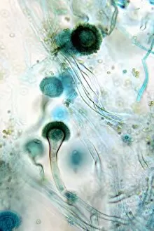
Hyphae, the intricate network of fungal filaments, are a fascinating subject to explore under the scanning electron microscope (SEM


All Professionally Made to Order for Quick Shipping
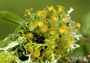
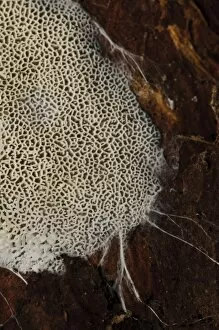
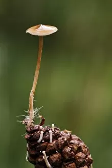
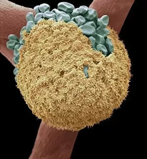
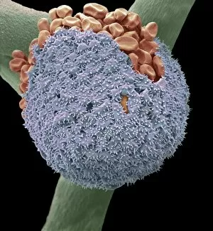
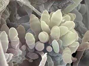
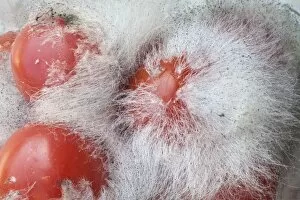
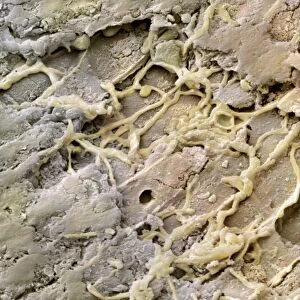
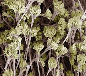
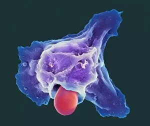
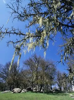
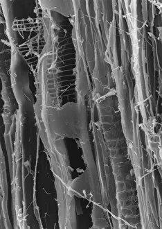
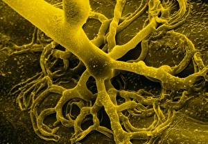
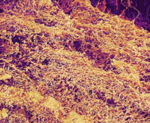
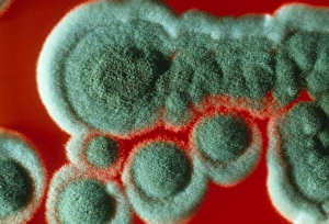
Hyphae, the intricate network of fungal filaments, are a fascinating subject to explore under the scanning electron microscope (SEM). From Candida fungus to lichen like Teloschistes chrysophthalmus, these microscopic structures reveal a world unseen by the naked eye. In one captivating SEM image, we witness mycorrhiza fungus entwined within plant roots. Magnified 700 times and printed on A4 paper, this image showcases the symbiotic relationship between fungi and plants in stunning detail. Moving on from plants, we delve into other habitats where hyphae thrive. Bread mould is captured in another SEM photograph, its delicate threads branching out across the surface. Rotten wood also becomes an enchanting sight as hyphae infiltrate its decaying fibers in yet another mesmerizing picture. Nature's ability to adapt and conquer is evident when observing a mouldy lemon through Picture No. 11675587. The intricate web covers every inch of this once vibrant fruit, reminding us of nature's relentless cycle of growth and decay. Sulphur Disco (Bisporella sulfurina) fruiting bodies emerge from dead wood with visible fungal hyphae at their base in Picture No. 11675586. This striking image highlights how fungi can transform lifeless matter into thriving ecosystems. Trechispora hymenocystis spreads over dead wood with fungal hyphae surrounding its margins at Clumber Park; it serves as a testament to nature's resilience even amidst death and decay. Lastly, we encounter Baeospora myosura growing from the tip of a pine cone with delicate hyphae encircling its base—a conifercone cap indeed. These images remind us that even in seemingly inhospitable environments such as pine cones or rotten wood, life finds a way through these remarkable networks called hyphae.