
Hypha Collection
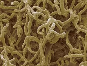
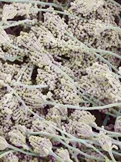
Hypha, the intricate network of thread-like structures found in fungi, plays a crucial role in their growth and survival


All Professionally Made to Order for Quick Shipping
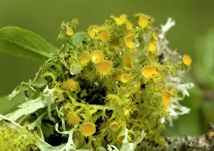
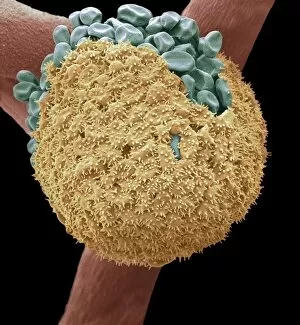
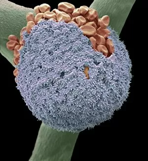
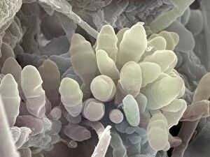
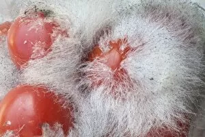
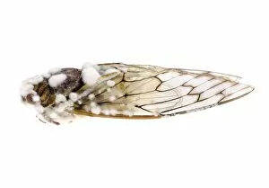
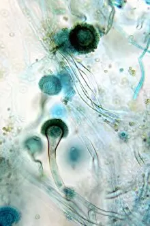
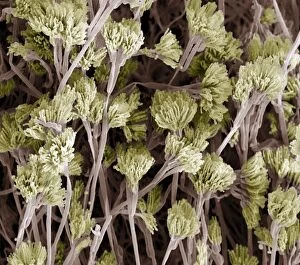
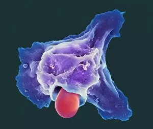
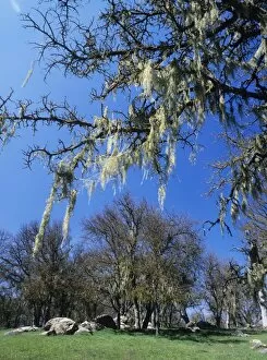
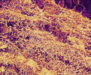
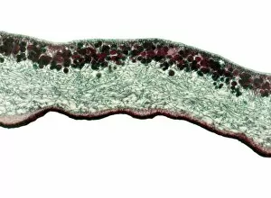
Hypha, the intricate network of thread-like structures found in fungi, plays a crucial role in their growth and survival. Candida fungus, known for causing infections in humans, showcases the complexity under scanning electron microscope (SEM). Similarly, lichen species like Teloschistes chrysophthalmus exhibit hyphal structures that aid them in nutrient absorption. In botany, understanding hypha is essential as it enables scientists to comprehend the fascinating world of fungi. Mycorrhizal fungus forms mutualistic associations with plant roots through its hyphal extensions, facilitating nutrient exchange between them. Illustrations depicting hypha within cells provide visual insights into this microscopic wonder. Bread mould and apple tree fungus captured under SEM further emphasize the diversity and intricacy of these fungal networks. The versatility is evident as bread mould (SEM C016 / 9052) and apple tree fungus (SEM C016 / 9417) display unique patterns and textures. Mould growing on tomatoes (C014 / 1426) exemplifies how certain conditions favor fungal growth while cicadas infected with fungus (C014 / 4582) demonstrate its impact on other organisms. Powdery mildew seen under SEM highlights the destructive potential of some fungal infections. Hypha's ability to invade host tissues can lead to diseases affecting various plants and animals alike. Exploring the world unveils an intricate web connecting different aspects of biology – from botany to mycology. Its significance lies not only in understanding fungi but also in comprehending ecological interactions shaped by these remarkable structures.