
Human Vertebra Collection
The human vertebra, also known as the backbone or spine, is a remarkable structure that provides support and protection to our bodies


All Professionally Made to Order for Quick Shipping
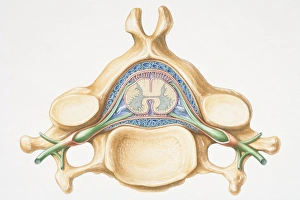
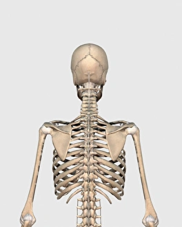
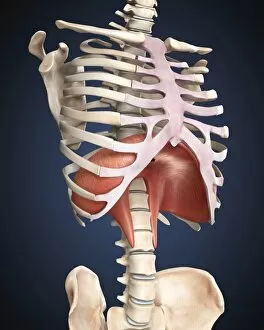
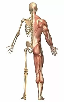
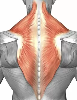
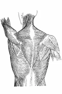
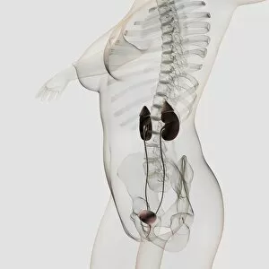
The human vertebra, also known as the backbone or spine, is a remarkable structure that provides support and protection to our bodies. This computer artwork showcases the intricate details of this essential part of our anatomy. Dating back to 1866, engravings such as the "Neck throat anatomy" and "Dorsal vertebrae anatomy" illustrations offer a glimpse into how early anatomists studied and understood the complexity of the human vertebra. One engraving from 1866 focuses on the "Cervical vertebrae, " highlighting their unique features in great detail. These bones play a crucial role in allowing us to move our heads freely and protect vital nerves within. From a rear view perspective, we can observe how these individual vertebral bones align to form the entire skeletal system's upper back, and is fascinating to see how each bone fits perfectly with its neighboring counterparts. Moving down towards the pelvis, an anterior view reveals this important region labeled for better understanding. The pelvis supports not only our weight but also plays a significant role in childbirth for women. Another intriguing visualization shows us the diaphragm - an essential muscle involved in breathing. Its position between the thoracic cavity and abdominal cavity allows it to contract and relax, aiding in respiration. The connection between skull and spine is depicted here, emphasizing their interdependence for proper functioning. The spinal cord passes through small openings within each vertebrae called intervertebral disks while being protected by ligaments surrounding them. A three-dimensional view highlights female sternum and rib cage structures; these are integral components that safeguard vital organs like heart and lungs while providing structural stability overall. Lastly, an anterior cross-section diagram demonstrates precisely how each vertebra fits around the spinal cord – showcasing both strength and flexibility simultaneously. These captivating images provide invaluable insights into human vertebral anatomy throughout history. They remind us of its significance in supporting our bodies' framework and protecting vital nerves, making it a truly remarkable part of our anatomy.