Fraud Blocker Skip to main content

Human Mouth Collection

The human mouth is a fascinating organ that serves multiple purposes

Background imageHuman Mouth Collection: Taste map of a human tongue, light green area sensitive to bitter taste

Taste map of a human tongue, light green area sensitive to bitter taste, yellow areas sensitive to sour taste, blue areas sensitive to salt taste, purple area sensitive to sweet taste

Background imageHuman Mouth Collection: Anatomy of Organs Engraving

Anatomy of Organs Engraving
Engraved illustrations of Anatomy of the Fasciae, Integuents, and Organs of Mastication and Respiration from Iconographic Encyclopedia of Science, Literature and Art, Published in 1851

Background imageHuman Mouth Collection: Maxilla bone with teeth

Maxilla bone with teeth
Illustration engraving of a maxilla bone with teeth

Background imageHuman Mouth Collection: Cross-section diagram of human mouth and jaw

Cross-section diagram of human mouth and jaw

Background imageHuman Mouth Collection: Illustration of human respiratory system showing oral cavity, and nasal cavity, larynx, trachea

Illustration of human respiratory system showing oral cavity, and nasal cavity, larynx, trachea, bronchus, and lungs

Background imageHuman Mouth Collection: Diagram illustrating the anatomy of the tongue, front view

Diagram illustrating the anatomy of the tongue, front view

Background imageHuman Mouth Collection: Anatomy of male respiratory system and internal organs

Anatomy of male respiratory system and internal organs
Anatomy of male respiratory system, including the mouth, cutaway view. Other internal organs are visible in half tones

Background imageHuman Mouth Collection: Anatomy of human mouth cavity

Anatomy of human mouth cavity

Background imageHuman Mouth Collection: Traditional Japanese Woodblock print of Actor

Traditional Japanese Woodblock print of Actor
This fine print by Utagawa Kuniyoshi 1797-1862 was one of the last great masters of the Japanese ukiyo-e style of woodblock prints he is associated with the Utagawa school

Background imageHuman Mouth Collection: Facial Nerves 19 century medical illustration

Facial Nerves 19 century medical illustration
Photograph of the original illustration from A System of Human Anatomy by Erasmus Wilson published in 1859

Background imageHuman Mouth Collection: Blonde Woman In Black Dress With Stripes & White Buttons & Trim Holding A Pilsner Glass

Blonde Woman In Black Dress With Stripes & White Buttons & Trim Holding A Pilsner Glass
UNITED STATES - Circa 1930s: Blonde Woman In Black Dress With Stripes & White Buttons & Trim Holding A Pilsner Glass Of Beer With A Large Head With Pursed Lips & Poppy Eyes Cartoon

Background imageHuman Mouth Collection: Woman with lips pursed and eyes wide open, portrait

Woman with lips pursed and eyes wide open, portrait
UNITED STATES - CIRCA 1940s: Woman with lips pursed and eyes wide open, portrait

Background imageHuman Mouth Collection: Cardiovascular System Engraving

Cardiovascular System Engraving
Engraved illustrations of The Vascular System from Iconographic Encyclopedia of Science, Literature and Art, Published in 1851. Copyright has expired on this artwork. Digitally restored

Background imageHuman Mouth Collection: Mouth wide open

Mouth wide open

Background imageHuman Mouth Collection: Diagram of nasal passages

Diagram of nasal passages

Background imageHuman Mouth Collection: Diagram showing inside of mouth and salivary glands

Diagram showing inside of mouth and salivary glands

Background imageHuman Mouth Collection: Black and white digital illustration of open mouth showing white teeth, tongue, and back of throat

Black and white digital illustration of open mouth showing white teeth, tongue, and back of throat

Background imageHuman Mouth Collection: Illustration of opened mouth with two rows of teeth showing

Illustration of opened mouth with two rows of teeth showing

Background imageHuman Mouth Collection: Anterior neck and facial muscles of the human head (with labels)

Anterior neck and facial muscles of the human head (with labels)

Background imageHuman Mouth Collection: Anatomy of human respiratory system

Anatomy of human respiratory system

Background imageHuman Mouth Collection: Human teeth structure with labels

Human teeth structure with labels

Background imageHuman Mouth Collection: Comparison of healthy gums versus gingivitis

Comparison of healthy gums versus gingivitis

Background imageHuman Mouth Collection: Anatomy of human face and neck muscles, front view

Anatomy of human face and neck muscles, front view

Background imageHuman Mouth Collection: View looking from inside the mouth while at the dentist

View looking from inside the mouth while at the dentist

Background imageHuman Mouth Collection: Cutaway diagram of human respiratory system

Cutaway diagram of human respiratory system, including nasal and mouth cross section

Background imageHuman Mouth Collection: Anatomy of a male human head, with half muscles and half skull

Anatomy of a male human head, with half muscles and half skull, front view

Background imageHuman Mouth Collection: Anatomy of human face muscles

Anatomy of human face muscles

Background imageHuman Mouth Collection: Cross section of human digestive system

Cross section of human digestive system

Background imageHuman Mouth Collection: Human digestive system, with labels

Human digestive system, with labels

Background imageHuman Mouth Collection: Blonde boy pouting, sticking out his lower lip, ready to cry

Blonde boy pouting, sticking out his lower lip, ready to cry
UNITED STATES - CIRCA 1950s: Blonde boy pouting, sticking out his lower lip, ready to cry

Background imageHuman Mouth Collection: aggression, anger, attitude, beaming, beauty, breaking through, breakthrough, brick

aggression, anger, attitude, beaming, beauty, breaking through, breakthrough, brick
Fierce woman kicking through brick wall

Background imageHuman Mouth Collection: Diagram of upper and lower teeth of a baby

Diagram of upper and lower teeth of a baby

Background imageHuman Mouth Collection: Illustration of open human vocal fold to inhale air, also known as vocal cords

Illustration of open human vocal fold to inhale air, also known as vocal cords

Background imageHuman Mouth Collection: Illustration of mouth showing healthy set of teeth

Illustration of mouth showing healthy set of teeth

Background imageHuman Mouth Collection: Human upper body showing bones, muscles and circulatory system

Human upper body showing bones, muscles and circulatory system

Background imageHuman Mouth Collection: Anatomy of inner ear and sinuses

Anatomy of inner ear and sinuses

Background imageHuman Mouth Collection: Human mouth open, showing teeth, gums and tongue

Human mouth open, showing teeth, gums and tongue
Medical illustration of human mouth open, showing teeth, gums and tongue

Background imageHuman Mouth Collection: Anatomy of human face muscles, side view

Anatomy of human face muscles, side view

Background imageHuman Mouth Collection: Smiling man with a spoon in the mouth, snacking

Smiling man with a spoon in the mouth, snacking

Background imageHuman Mouth Collection: Tablet, pill on a tongue, mouth

Tablet, pill on a tongue, mouth

Background imageHuman Mouth Collection: Young, desperate man wearing a shirt and a tie holding his hands over his ears

Young, desperate man wearing a shirt and a tie holding his hands over his ears, has 100-euro notes stuffed into his mouth

Background imageHuman Mouth Collection: Young woman in black dress with fabric roses, portrait

Young woman in black dress with fabric roses, portrait

Background imageHuman Mouth Collection: Baby girl (9-12 months) sitting in high chair, holding finger in mouth, (B&W)

Baby girl (9-12 months) sitting in high chair, holding finger in mouth, (B&W)

Background imageHuman Mouth Collection: Human speech organs, wood engraving, published in 1880

Human speech organs, wood engraving, published in 1880
The Organs of Speech: A) mouth, B) uvula, C) tongue, D) tonsils, E) epiglottis, F) thyroid cartilage, G) arytenoid muscle, H+I) vestibular folds, K) vocal fold, L) larynx, M) hypopharynx, N) trachea

Background imageHuman Mouth Collection: Baby sitting in high chair, sticking out tongue. (Photo by H)

Baby sitting in high chair, sticking out tongue. (Photo by H. Armstrong Roberts / Retrofile / Getty Images)
UNITED STATES - CIRCA 1930s: Baby sitting in high chair, sticking out tongue

Background imageHuman Mouth Collection: beauty, bonding, cloud, color image, computer graphic, concept, covering, cyborg

beauty, bonding, cloud, color image, computer graphic, concept, covering, cyborg
" Woman robots covering eyes, ears and mouth"

Background imageHuman Mouth Collection: Ophioglossum vulgatum (adder s-tongue)

Ophioglossum vulgatum (adder s-tongue)
Antique illustration of Ophioglossum vulgatum (adder s-tongue)


All Professionally Made to Order for Quick Shipping

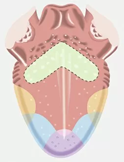
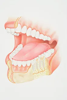
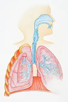
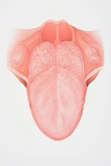
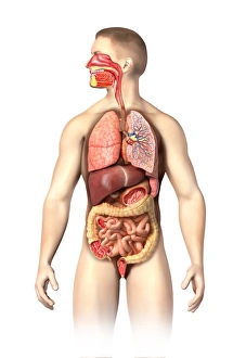
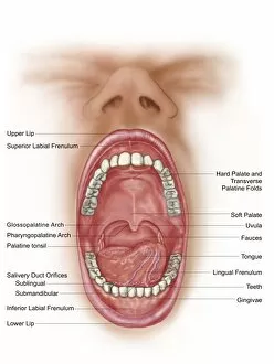
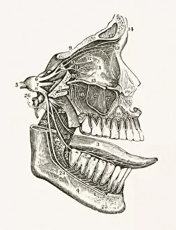
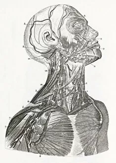
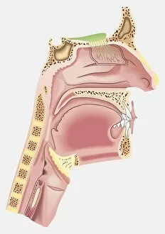
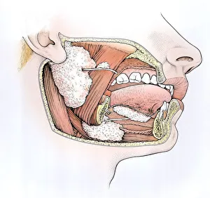
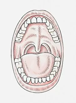
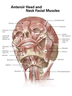
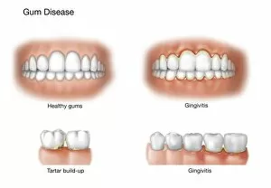
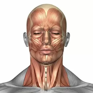
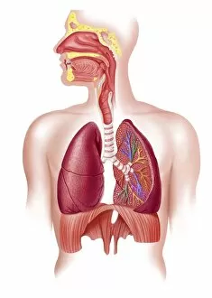
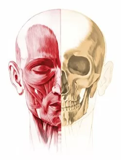
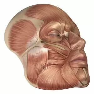
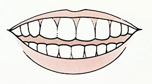
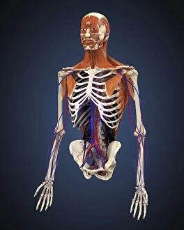
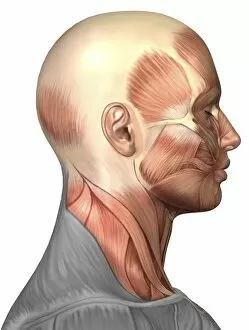
The human mouth is a fascinating organ that serves multiple purposes. It not only allows us to communicate through speech but also plays a crucial role in our sense of taste and the process of digestion. Did you know that the tongue has different areas sensitive to various tastes? According to the taste map, the light green area on your tongue is particularly sensitive to bitter tastes, while yellow areas detect sour tastes. The blue regions are responsible for sensing saltiness, and the purple area is where sweet flavors are detected. This intricate system helps us savor and differentiate between various flavors. When we examine the anatomy of the mouth, we can see its complexity. From an engraving depicting organs, we observe how it connects with other vital systems like respiration and digestion. The cross-section diagram showcases both our oral cavity and jaw structure, highlighting their interdependence. Speaking of which, let's not forget about teeth. A maxilla bone illustration shows how these essential tools for chewing are anchored within our mouths. They play a significant role in breaking down food before it enters our digestive system. Furthermore, understanding respiratory functions is crucial when studying the human mouth cavity. An illustration displaying nasal cavities, larynx, trachea, bronchus, and lungs demonstrates how air passes through this pathway during breathing. Now let's focus solely on one aspect: the tongue itself. A front view diagram reveals its anatomical details—taste buds covering its surface enable us to perceive different flavors accurately. Moving beyond scientific depictions into artistry; a traditional Japanese woodblock print captures an actor's expressive face—an embodiment of emotions conveyed through facial features including lips that shape words or express feelings such as pouting or crying—a testament to how much power lies within this small part of our body. Lastly, exploring medical illustrations from centuries ago reminds us of historical knowledge gained about facial nerves' intricacies related to sensation around our mouths—a reminder that even long ago, humans were fascinated by this remarkable organ.