
Human Brain Collection (page 3)
"The intricate web of the human brain unraveled: Exploring its anatomy and functions" Delve into the depths of the human brain, as seen from an inferior view


All Professionally Made to Order for Quick Shipping
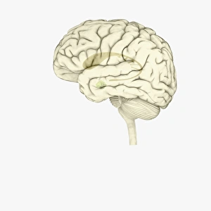
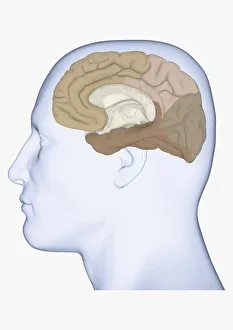
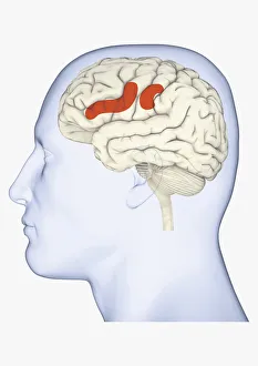
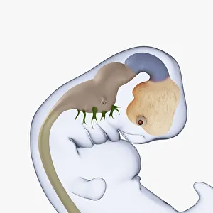
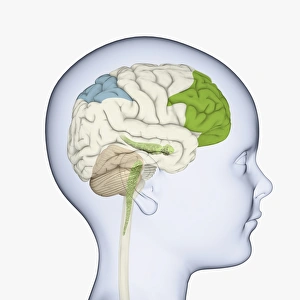
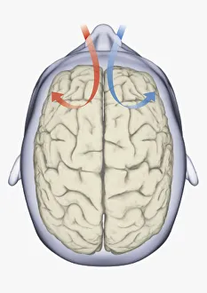
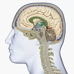
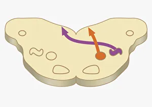
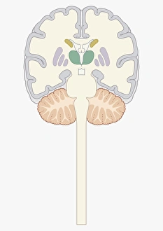
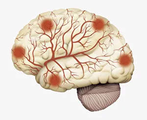
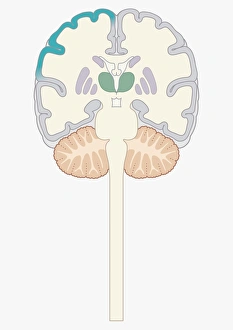
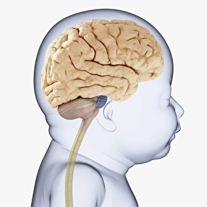
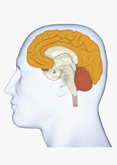
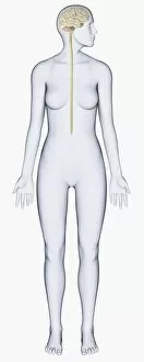
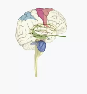
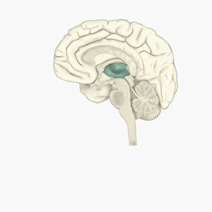
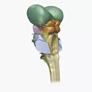
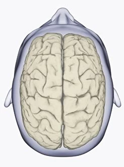
"The intricate web of the human brain unraveled: Exploring its anatomy and functions" Delve into the depths of the human brain, as seen from an inferior view, revealing its complex structure. White matter fibres intricately interconnect regions of this remarkable organ, forming a vast network that facilitates communication. Step back in time with artwork from 1930 (C015) and an engraving dating back to 1895, showcasing early attempts to understand the brain's inner workings. Cross-section illustrations unveil the limbic system and primitive forebrain, shedding light on our emotional responses and primal instincts. A superior view presents a colorful tapestry of lobes labeled with precision – a visual representation of different areas responsible for various cognitive processes. From perception to language comprehension, each lobe plays a vital role in shaping our thoughts and actions. Observe the lateral view anatomy; it reveals hidden secrets within its convoluted folds. The white matter fibres (C014 / 5666) form intricate pathways connecting distant regions, enabling seamless information transfer throughout this neural powerhouse. Travel further back in time with an engraved illustration circa 1880 depicting the under surface of this enigmatic organ. A testament to our ancestors' curiosity about what lies beneath consciousness itself. Witness how modern technology has revolutionized our understanding through digital illustrations portraying connections between the nervous system, spinal cord, and brain. It highlights their inseparable bond - orchestrating every movement we make or sensation we feel. Explore yet another glimpse into white matter fibres (C014 / 5668), reinforcing their significance in transmitting signals across vast distances within this cerebral universe. As we unravel these mysteries surrounding the human brain's complexity and beauty, let us not forget those affected by dementia—a condition that challenges memory retention but never diminishes humanity's resilience or spirit for discovery. In awe-inspiring fashion akin to a rocket launch into space—our brains propel us into the realms of knowledge, creativity, and innovation.