Fraud Blocker Skip to main content

Human Body Part Collection (page 3)

The intricate beauty of our human body never ceases to amaze

Background imageHuman Body Part Collection: Portrait of a young man, cool tone

Portrait of a young man, cool tone

Background imageHuman Body Part Collection: Portrait of a young man in sunlight, beauty shot

Portrait of a young man in sunlight, beauty shot

Background imageHuman Body Part Collection: Young man wearing bathing trunks, leaning against a concrete wall

Young man wearing bathing trunks, leaning against a concrete wall

Background imageHuman Body Part Collection: Sensual face of a young woman, close-up

Sensual face of a young woman, close-up

Background imageHuman Body Part Collection: Girl, teenager, serious

Girl, teenager, serious

Background imageHuman Body Part Collection: Young man with laptop sitting on steps

Young man with laptop sitting on steps

Background imageHuman Body Part Collection: Knee joint

Knee joint with cruciate ligaments

Background imageHuman Body Part Collection: Digital illustration of dilated pupil of human eye with contraction of outer radial muscle fibres

Digital illustration of dilated pupil of human eye with contraction of outer radial muscle fibres

Background imageHuman Body Part Collection: Illustration showing diaphragm moving down when exhaling and up when inhaling

Illustration showing diaphragm moving down when exhaling and up when inhaling

Background imageHuman Body Part Collection: Buddha head in tree roots, Wat Mahathat, Ayutthaya

Buddha head in tree roots, Wat Mahathat, Ayutthaya
The location of this site is Wat Mahathat in Ayutthaya. This ancient temple was built during the 14th century, but was reduced to ruins in 1767 when the Burmese army invaded Ayutthaya

Background imageHuman Body Part Collection: Illustration of hands on synthesizer keyboard

Illustration of hands on synthesizer keyboard

Background imageHuman Body Part Collection: Young woman, 25, holding a sunflower in front of her face

Young woman, 25, holding a sunflower in front of her face

Background imageHuman Body Part Collection: Crystal skull and Maya pyramid, artwork

Crystal skull and Maya pyramid, artwork
Crystal skull and Maya pyramid, computer artwork

Background imageHuman Body Part Collection: Crying baby boy (15-18 months) in baby seat, (B&W), close-up

Crying baby boy (15-18 months) in baby seat, (B&W), close-up

Background imageHuman Body Part Collection: Molar tooth

Molar tooth
Tooth, transparent cross section of a molar tooth with arteries (red), veins (purple) and nerves (green)

Background imageHuman Body Part Collection: Total knee replacement, X-rays

Total knee replacement, X-rays
Total knee replacement. Coloured frontal (left) and profile (right) X-rays of the right knee of a 69 year old patient after total knee replacement surgery

Background imageHuman Body Part Collection: The muscles of the human abdominal wall

The muscles of the human abdominal wall

Background imageHuman Body Part Collection: Normal lateral view of the lumbar vertebrae showing spinal nerve roots

Normal lateral view of the lumbar vertebrae showing spinal nerve roots
Detail, Anatomy, Diagram, Illustration, Spine, Bone, Medical, Biology, Disk, Normal, Cut Out, White Background, Vertical, Artwork, Color Image, Close Up, Healthcare And Medicine, The Human Body

Background imageHuman Body Part Collection: Front view of the female anatomy hilighting the endocrine system

Front view of the female anatomy hilighting the endocrine system
Brain, Anatomy, Diagram, Heart, Illustration, Bone, Shoulder, Medical, Uterus, Biology, Pelvis, Femur, See Through, Vertical, Artwork, Female Likeness, Color Image, Front View

Background imageHuman Body Part Collection: William Buckland (1784-1856)

William Buckland (1784-1856)
863927 William Buckland (1784-1856); (add.info.: Sectional view of a bone cavern showing debris of animal bones. William Buckland (1784-1856) English geologist and clergyman

Background imageHuman Body Part Collection: Posterior view of a normal heart and its arteries

Posterior view of a normal heart and its arteries
Detail, Anatomy, Diagram, Muscle, Heart, Illustration, Medical, Biology, Cardiac, Rear View, Cardiovascular, Cut Out, White Background, Vertical, Artwork, Color Image, Close Up

Background imageHuman Body Part Collection: Normal cross section of the skin in layers

Normal cross section of the skin in layers
Detail, Anatomy, Diagram, Illustration, Layer, Medical, Vein, Skin, Biology, Fat, Veins, Epidermis, Cut Out, White Background, Hair, Vertical, Artwork, Cross Section, Color Image, Close Up

Background imageHuman Body Part Collection: Skin cross section showing a blackhead

Skin cross section showing a blackhead
Detail, Anatomy, Diagram, Illustration, Medical, Vein, Skin, Biology, Fat, Veins, Epidermis, Cut Out, White Background, Hair, Vertical, Artwork, Cross Section, Color Image, Close Up

Background imageHuman Body Part Collection: Anterior view of pelvis with hip bones showing arthritis and osteophytes on femoral heads

Anterior view of pelvis with hip bones showing arthritis and osteophytes on femoral heads
Horizontal, Anatomy, Diagram, Illustration, Bone, Disease, Medical, Hip, Biology, Pelvis, Femur, Cut Out, Arthritis, White Background, Artwork, Color Image, Front View, Healthcare And Medicine

Background imageHuman Body Part Collection: Normal anatomy of the eye in cross section

Normal anatomy of the eye in cross section
Horizontal, Vision, Detail, Anatomy, Diagram, Muscle, Illustration, Lens, Iris, Medical, Eyeball, Biology, Lateral, Cut Out, White Background, Eye, Artwork, Cross Section, Color Image, Close Up

Background imageHuman Body Part Collection: Normal front view of the adult skull

Normal front view of the adult skull
Anatomy, Diagram, Illustration, Bone, Teeth, Skull, Medical, Biology, Cut Out, White Background, Vertical, Artwork, Color Image, Front View, Human Skull, Healthcare And Medicine, The Human Body

Background imageHuman Body Part Collection: Normal anterior view of pelvis with hip bones

Normal anterior view of pelvis with hip bones
Horizontal, Anatomy, Diagram, Illustration, Bone, Medical, Hip, Biology, Pelvis, Femur, Cut Out, White Background, Artwork, Color Image, Front View, Healthcare And Medicine, The Human Body

Background imageHuman Body Part Collection: Showing a proper way to cut toe nails versus and improper way, shown as a rounded cut

Showing a proper way to cut toe nails versus and improper way, shown as a rounded cut
Horizontal, Detail, Anatomy, Diagram, Illustration, Toe, Advice, Text, Biology, Toenail, Blue Background, Contrasts, Artwork, Color Image, Close Up, Human Representation, Healthcare And Medicine

Background imageHuman Body Part Collection: Lateral view of a normal brain

Lateral view of a normal brain
Horizontal, Brain, Detail, Anatomy, Diagram, Illustration, Pink, Medical, Biology, Cut Out, White Background, Artwork, Color Image, Close Up, Healthcare And Medicine, The Human Body, No People

Background imageHuman Body Part Collection: Front view of a woman nine months pregnant (baby phantomed within) ready for delivery

Front view of a woman nine months pregnant (baby phantomed within) ready for delivery
Detail, Baby, Anatomy, Diagram, Mother, Illustration, Bone, Birth, Medical, Fetus, Pregnant, Pregnancy, Biology, Pelvis, Femur, Development, Abdomen, Anterior, See Through, Cut Out, White Background

Background imageHuman Body Part Collection: Repetitive activities like aerobics that involve lifting up the knee can cause trauma to

Repetitive activities like aerobics that involve lifting up the knee can cause trauma to
Detail, Anatomy, Diagram, Muscle, Pain, Illustration, Bone, Trauma, Injury, Medical, Biology, Pelvis, Femur, See Through, Cut Out, White Background, Artwork, Color Image, Front View, Close Up

Background imageHuman Body Part Collection: Illustration of dorsal wrist Ganglion cyst

Illustration of dorsal wrist Ganglion cyst
Detail, Anatomy, Diagram, Pain, Illustration, Bone, Injury, Medical, Biology, Wrist, See Through, Cut Out, White Background, Vertical, Artwork, Color Image, Close Up, Healthcare And Medicine

Background imageHuman Body Part Collection: Snapping hip syndrome occurs when the iliopsoas tendon subluxes over the greater

Snapping hip syndrome occurs when the iliopsoas tendon subluxes over the greater
Horizontal, Anatomy, Diagram, Muscle, Illustration, Bone, Medical, Hip, Biology, Pelvis, Femur, See Through, Rear View, Cut Out, Arthritis, White Background, Artwork, Color Image, Front View

Background imageHuman Body Part Collection: Yoga Sun Salutations Sequence Illustration

Yoga Sun Salutations Sequence Illustration
Illustration Steps of Yoga Sun Salutation

Background imageHuman Body Part Collection: Labelled Human Skeleton. Engraving

Labelled Human Skeleton. Engraving
Labelled engraved image of human skeleton

Background imageHuman Body Part Collection: Joint pain, conceptual artwork

Joint pain, conceptual artwork
Joint pain, conceptual computer artwork

Background imageHuman Body Part Collection: Healthy ankle joint, X-ray

Healthy ankle joint, X-ray
Healthy ankle joint. Coloured profile X-ray of the left ankle of a 21 year old patient. Strapping around the ankle is visible on this X-ray

Background imageHuman Body Part Collection: Scoliosis of the spine, X-ray

Scoliosis of the spine, X-ray
Scoliosis of the spine. Coloured X-ray of the chest of a 31 year old patient with scoliosis (sideways curvature) of the spine

Background imageHuman Body Part Collection: Rheumatoid arthritis, X-ray

Rheumatoid arthritis, X-ray
Rheumatoid arthritis. Coloured X-ray of the hands of an 81 year old female patient with rheumatoid arthritis

Background imageHuman Body Part Collection: Normal lower leg, X-ray

Normal lower leg, X-ray
Normal lower leg. X-ray of the lower leg of a 24 year old woman

Background imageHuman Body Part Collection: Normal abdomen, X-ray

Normal abdomen, X-ray
Normal abdomen. X-ray of the abdomen of a 20 year old female

Background imageHuman Body Part Collection: Section of the Lungs Anatomy Drawings 1888

Section of the Lungs Anatomy Drawings 1888
Illustrated Natural History of the Three Kingdoms Illustrierte Naturgeschichte der drei Reiche Franz StrAÔé¼ssles - Wilhelm Nitzschke, Stuttgart 1888

Background imageHuman Body Part Collection: Human skull anatomy illustration 1866

Human skull anatomy illustration 1866
Atlas d anatomie descriptive du corps humain

Background imageHuman Body Part Collection: Bones of hand anatomy engraving 1857

Bones of hand anatomy engraving 1857
Rank, johannes - The human being. 1 - 1894

Background imageHuman Body Part Collection: Skull

Skull
Engraving From 1898 Featuring A Human Skull


All Professionally Made to Order for Quick Shipping

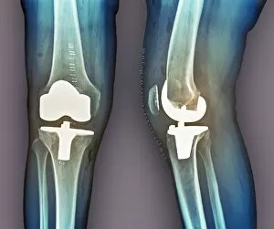
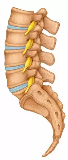
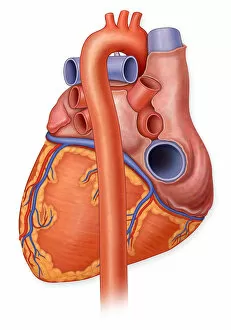
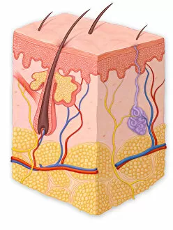
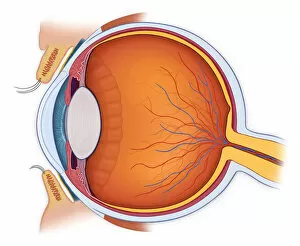
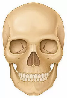
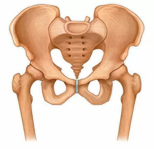
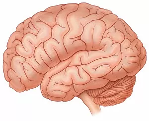
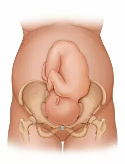
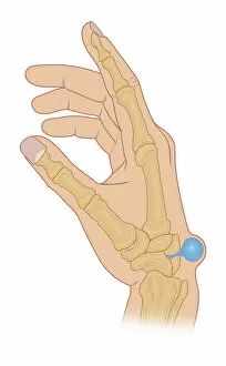
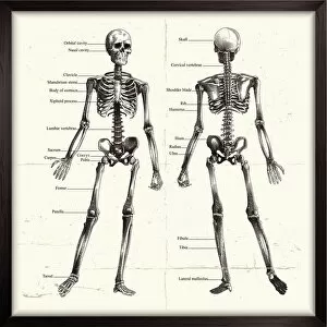
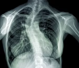
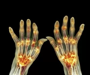
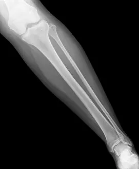
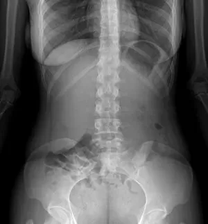
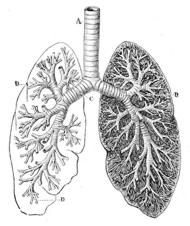
The intricate beauty of our human body never ceases to amaze. From the normal knees that allow us to walk and run, to the X-ray revealing their hidden structure, every part tells a unique story. Just like Janus, the Roman god with two faces looking towards the past and future, our brain anatomy engraving from 1895 reminds us of our complex thoughts and emotions. As we explore further, an illustration of the female lymphatic system unveils a network vital for immune function. The Human Heart Anatomy from 1888 showcases its remarkable design responsible for keeping us alive with each beat. Moving upwards, a diagram of facial muscles reveals how expressions shape our interactions. Communication takes various forms; even sign language has its place in expressing ourselves. An illustration displaying 26 hand signals representing letters reminds us of this beautiful diversity. But wait. Santa Claus at the beach? Well, sometimes even iconic figures need some relaxation too. Returning to our focus on brains, MRI scans provide glimpses into their inner workings while a cross-section illustration highlights both primitive instincts and emotional responses within the limbic system. Amidst all these scientific wonders lies humor – captured by a man smashing cake on another's face – reminding us not to take life too seriously. Finally, let's not forget about our lymphatic system once more as it plays an essential role in maintaining overall health. Whether through art or science, these captivating images celebrate different aspects of humanity – from physical structures that enable movement and expression to intricate systems that keep us alive.