
Hip Joint Collection
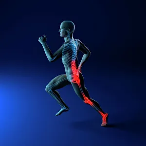
The hip joint is a complex structure that plays a crucial role in our mobility and overall well-being and can be susceptible to various running injuries


All Professionally Made to Order for Quick Shipping
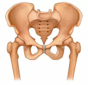
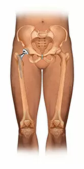
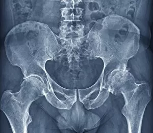
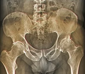
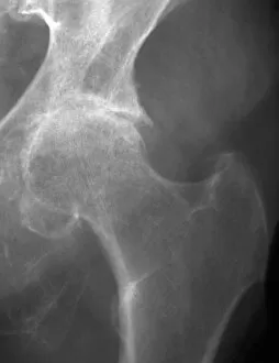
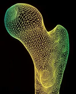
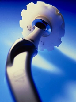
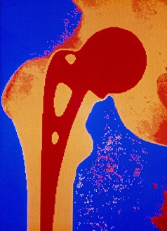
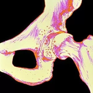
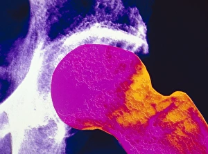
The hip joint is a complex structure that plays a crucial role in our mobility and overall well-being and can be susceptible to various running injuries, which can cause discomfort and hinder our ability to stay active. In the world of conceptual artwork, the hip joint has been depicted in fascinating ways, showcasing its intricate design and importance. Osteoarthritis of the hip is a condition that affects many individuals, causing pain and stiffness in the joint. X-ray images reveal this degenerative disease, with visible signs such as arthritis and osteophytes on the femoral heads. A normal anterior view of the pelvis with hip bones shows us how this joint should ideally look like - healthy and functional. However, when affected by osteoarthritis, an anterior view reveals significant changes due to damage caused by wear-and-tear over time. For those who have undergone total hip replacement surgery, a front view of their body proudly displays their new lease on life - thanks to modern medical advancements. Illustrations depicting human hip-joints provide valuable insights into its anatomy and functioning mechanisms. These visuals help us understand better how this remarkable joint enables movement while maintaining stability. Zooming in on a close-up image of a hip joint prosthesis highlights the intricacy involved in creating artificial joints for those who need them most. X-rays labeled F006/3744, F006/3745, C018/0593 showcase different cases of osteoarthritis affecting the hips. Each image tells its own story about how this condition impacts individuals differently but shares common symptoms such as pain and reduced range of motion. Even ancient beings like Australopithecus sp. , known for their bipedal locomotion capabilities similar to humans', had unique features within their thigh & hip bones that give us insight into our evolutionary history.