Fraud Blocker Skip to main content

Hematological Collection

"Hematological Wonders: Exploring the Intricacies of Blood and Beyond" Delving into the microscopic world

Background imageHematological Collection: White blood cells and platelets, SEM C016 / 3099

White blood cells and platelets, SEM C016 / 3099
White blood cells and platelets. Coloured scanning electron micrograph (SEM) of platelets (green) with a variety of different white blood cells (leucocytes, yellow)

Background imageHematological Collection: White blood cells and platelets, SEM C016 / 3098

White blood cells and platelets, SEM C016 / 3098
White blood cells and platelets. Coloured scanning electron micrograph (SEM) of platelets (purple) with a variety of different white blood cells (leucocytes, blue)

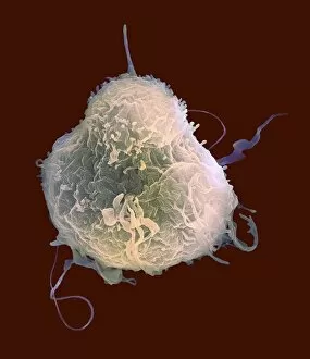
Background imageHematological Collection: Monocyte white blood cell, SEM C016 / 3089

Monocyte white blood cell, SEM C016 / 3089
Monocyte white blood cell. Scanning electron micrograph (SEM) of a monocyte white blood cell. Monocytes, like all white blood cells, are part of the human bodys immune system

Background imageHematological Collection: Red blood cells, light micrograph C016 / 3035

Red blood cells, light micrograph C016 / 3035
Red blood cells. Differential interference contrast (DIC) micrograph of red blood cells (erythrocytes). Red blood cells are biconcave, giving them a large surface area for gas exchange

Background imageHematological Collection: Thrombosed blood vessel, artwork C013 / 4649

Thrombosed blood vessel, artwork C013 / 4649
Thrombosed blood vessel. Computer artwork of a blood clot (thrombus) formed on the internal wall of a blood vessel

Background imageHematological Collection: Blood samples C018 / 1034

Blood samples C018 / 1034
Blood samples

Background imageHematological Collection: Blood sample F006 / 9054

Blood sample F006 / 9054
Blood sample and syringe

Background imageHematological Collection: Blood sample F006 / 9055

Blood sample F006 / 9055
Blood sample

Background imageHematological Collection: Haematopoietic stem cells, artwork

Haematopoietic stem cells, artwork
Haematopoietic stem cells. Cutaway computer artwork showing white blood cells (leucocytes, white, round), red blood cells (erythrocytes, red) and haematopoietic stem cells (HSCs)

Background imageHematological Collection: Thickened skull, MRI scan

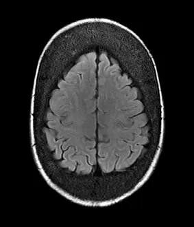
Thickened skull, MRI scan
Thickened skull. Magnetic resonance imaging (MRI) scan of an axial section through the head of a 22-year-old patient, showing thickening of the trabecular (spongy) bone of the skull

Background imageHematological Collection: Atherosclerosis, 3D CT scan

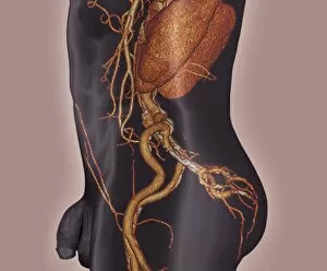
Atherosclerosis, 3D CT scan
Atherosclerosis. 3D computed tomography (CT) angiogram of the main blood vessels of the lower body a 58-year-old man, showing numerous atheromatous plaques (light, centre) on the vessel walls

Background imageHematological Collection: White blood cell antigen presentation C016 / 9058

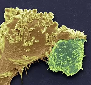
White blood cell antigen presentation C016 / 9058
White blood cell antigen presentation. Coloured scanning electron micrograph (SEM) showing the interaction between a macrophage (yellow) and a T helper lymphocyte (Th cell, green)

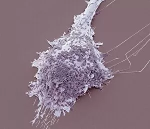
Background imageHematological Collection: Microglial white blood cell, SEM C016 / 9115

Microglial white blood cell, SEM C016 / 9115
Microglial white blood cell. Coloured scanning electron micrograph (SEM) of a microglial cell. Microglia are phagocytic macrophage white blood cells that are the main form of immune system in

Background imageHematological Collection: Light microscopy C016 / 9700

Light microscopy C016 / 9700
Light microscopy. Light microscope being used to magnify a blood sample

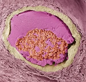
Background imageHematological Collection: Blood clot, SEM C016 / 9088

Blood clot, SEM C016 / 9088
Blood clot. Coloured scanning electron micrograph (SEM) of a section through a blood clot (thrombus) in an arteriole (small blood vessel), showing red blood cells (red) in a fibrin mesh (green)

Background imageHematological Collection: Microglial white blood cell, SEM C016 / 9119

Microglial white blood cell, SEM C016 / 9119
Microglial white blood cell. Coloured scanning electron micrograph (SEM) of a microglial cell. Microglia are phagocytic macrophage white blood cells that are the main form of immune system in

Background imageHematological Collection: Microglial white blood cell, SEM C016 / 9117

Microglial white blood cell, SEM C016 / 9117
Microglial white blood cell. Coloured scanning electron micrograph (SEM) of a microglial cell. Microglia are phagocytic macrophage white blood cells that are the main form of immune system in

Background imageHematological Collection: Platelet, SEM C016 / 9060

Platelet, SEM C016 / 9060
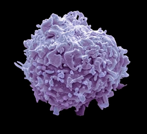
Platelet, coloured scanning electron micrograph (SEM). Platelets, or thrombocytes, are fragments of white blood cells. When blood clots

Background imageHematological Collection: Lymphocyte white blood cell, SEM C016 / 9414

Lymphocyte white blood cell, SEM C016 / 9414
Lymphocyte white blood cell. Coloured scanning electron micrograph (SEM) of a lymphocyte white blood cell. Lymphocytes are involved in the immune systems defence mechanisms, lymph system

Background imageHematological Collection: Microglial white blood cell, SEM C016 / 9109

Microglial white blood cell, SEM C016 / 9109
Microglial white blood cell. Coloured scanning electron micrograph (SEM) of a microglial cell. Microglia are phagocytic macrophage white blood cells that are the main form of immune system in

Background imageHematological Collection: Light microscopy C016 / 9703

Light microscopy C016 / 9703
Light microscopy. Light microscope being used to magnify a blood sample

Background imageHematological Collection: Crenated red blood cells, SEM C016 / 9029

Crenated red blood cells, SEM C016 / 9029
Crenated red blood cells. Coloured scanning electron micrograph (SEM) of a section through an arteriole (small blood vessel), showing crenated red blood cells inside

Background imageHematological Collection: Human bone structure, artwork C016 / 7494

Human bone structure, artwork C016 / 7494
Human bone structure. Computer artwork showing all the different parts that make up a human bone, including the bone marrow (bottom left) where blood cells (round, white and red) are formed

Background imageHematological Collection: Blood clot, SEM C016 / 9089

Blood clot, SEM C016 / 9089
Blood clot. Coloured scanning electron micrograph (SEM) of a section through a blood clot (thrombus) in an arteriole (small blood vessel), showing red blood cells (red) in a fibrin mesh (bright pink)

Background imageHematological Collection: Lymphocyte white blood cell, SEM C016 / 9415

Lymphocyte white blood cell, SEM C016 / 9415
Lymphocyte white blood cell. Coloured scanning electron micrograph (SEM) of a lymphocyte white blood cell. Lymphocytes are involved in the immune systems defence mechanisms, lymph system

Background imageHematological Collection: Coronary blood clot, SEM C016 / 9133

Coronary blood clot, SEM C016 / 9133
Coronary blood clot. Coloured scanning electron micrograph (SEM) of a blood clot (thrombus) in the coronary artery, showing red blood cells (purple) in a fibrin mesh (threads)

Background imageHematological Collection: Lymphocyte white blood cells, SEM C016 / 9140

Lymphocyte white blood cells, SEM C016 / 9140
Lymphocyte white blood cells. Coloured scanning electron micrograph (SEM) of lymphocyte white blood cells from peripheral blood

Background imageHematological Collection: White blood cell antigen presentation C016 / 9057

White blood cell antigen presentation C016 / 9057
White blood cell antigen presentation. Coloured scanning electron micrograph (SEM) showing the interaction between a macrophage (red) and a T helper lymphocyte (Th cell, yellow)

Background imageHematological Collection: Microglial white blood cell, SEM C016 / 9114

Microglial white blood cell, SEM C016 / 9114
Microglial white blood cell. Coloured scanning electron micrograph (SEM) of a microglial cell. Microglia are phagocytic macrophage white blood cells that are the main form of immune system in

Background imageHematological Collection: Microglial white blood cells, SEM C016 / 9113

Microglial white blood cells, SEM C016 / 9113
Microglial white blood cells. Coloured scanning electron micrograph (SEM) of microglial cells. Microglia are phagocytic macrophage white blood cells that are the main form of immune system in

Background imageHematological Collection: Cyanide and strychnine in blood, artwork C016 / 7682

Cyanide and strychnine in blood, artwork C016 / 7682
Cyanide and strychnine in blood. Computer artwork showing the structural formulas of cyanide and strychnine in blood (red)

Background imageHematological Collection: Microglial white blood cell, SEM C016 / 9118

Microglial white blood cell, SEM C016 / 9118
Microglial white blood cell. Coloured scanning electron micrograph (SEM) of a microglial cell. Microglia are phagocytic macrophage white blood cells that are the main form of immune system in

Background imageHematological Collection: Light microscopy C016 / 9702

Light microscopy C016 / 9702
Light microscopy. Light microscope being used to magnify a blood sample

Background imageHematological Collection: Microglial white blood cells, SEM C016 / 9111

Microglial white blood cells, SEM C016 / 9111
Microglial white blood cells. Coloured scanning electron micrograph (SEM) of microglial cells. Microglia are phagocytic macrophage white blood cells that are the main form of immune system in

Background imageHematological Collection: Platelet, SEM C016 / 9059

Platelet, SEM C016 / 9059
Platelet, coloured scanning electron micrograph (SEM). Platelets, or thrombocytes, are fragments of white blood cells. When blood clots

Background imageHematological Collection: Microglial white blood cells, SEM C016 / 9112

Microglial white blood cells, SEM C016 / 9112
Microglial white blood cells. Coloured scanning electron micrograph (SEM) of microglial cells. Microglia are phagocytic macrophage white blood cells that are the main form of immune system in

Background imageHematological Collection: Microglial white blood cell, SEM C016 / 9116

Microglial white blood cell, SEM C016 / 9116
Microglial white blood cell. Coloured scanning electron micrograph (SEM) of a microglial cell. Microglia are phagocytic macrophage white blood cells that are the main form of immune system in

Background imageHematological Collection: Lymphocyte white blood cells, SEM C016 / 9138

Lymphocyte white blood cells, SEM C016 / 9138
Lymphocyte white blood cells. Coloured scanning electron micrograph (SEM) of lymphocyte white blood cells from peripheral blood

Background imageHematological Collection: Light microscopy C016 / 9701

Light microscopy C016 / 9701
Light microscopy. Light microscope being used to magnify a blood sample

Background imageHematological Collection: Macrophage white blood cells, TEM

Macrophage white blood cells, TEM
Macrophage white blood cells. Transmission electron micrograph (TEM) of a section through two macrophages located within connective tissue, showing the nuclei and numerous lysosomes (dark spots)

Background imageHematological Collection: Leukaemia cell, SEM

Leukaemia cell, SEM
Leukaemia cell. Coloured scanning electron micrograph (SEM) of a leukaemic (cancerous) lymphocyte white blood cell (green), amongst normal red blood cells (erythrocytes, red)

Background imageHematological Collection: Antibodies and B cells, artwork C016 / 6255

Antibodies and B cells, artwork C016 / 6255
Antibodies and B cells. Computer artwork showing antibodies (green) neutralising cytokine activity in B cell (large, round) white blood cells

Background imageHematological Collection: Antibodies and B cells, artwork C016 / 6254

Antibodies and B cells, artwork C016 / 6254
Antibodies and B cells. Computer artwork showing antibodies (green) neutralising cytokine activity in B cell (large, round) white blood cells

Background imageHematological Collection: Normal and leukaemic blood, artwork C016 / 6261

Normal and leukaemic blood, artwork C016 / 6261
Normal and leukaemic blood. Computer artwork comparing the appearance of normal blood (left) and that affected by leukaemia (right)

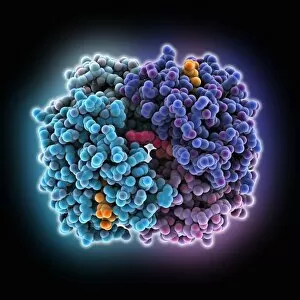
Background imageHematological Collection: Haemoglobin, molecular model C015 / 8938

Haemoglobin, molecular model C015 / 8938
Haemoglobin, molecular model. This molecule transports oxygen around the body in red blood cells. It consists of four globin proteins (amino acid chains; orange, green, blue and purple)

Background imageHematological Collection: Haemoglobin, molecular model C015 / 9391

Haemoglobin, molecular model C015 / 9391
Haemoglobin, molecular model. This is deoxyhaemoglobin, the molecule in its non-oxygen bound state. Haemoglobin transports oxygen around the body in red blood cells

Background imageHematological Collection: Haemoglobin, molecular model C015 / 8939

Haemoglobin, molecular model C015 / 8939
Haemoglobin, molecular model. This molecule transports oxygen around the body in red blood cells. It consists of four globin proteins (amino acid chains; orange, green, blue and purple)

Background imageHematological Collection: Haemoglobin, molecular model C015 / 9392

Haemoglobin, molecular model C015 / 9392
Haemoglobin, molecular model. This is deoxyhaemoglobin, the molecule in its non-oxygen bound state. Haemoglobin transports oxygen around the body in red blood cells


All Professionally Made to Order for Quick Shipping

"Hematological Wonders: Exploring the Intricacies of Blood and Beyond" Delving into the microscopic world, we witness the intricate dance of white blood cells and platelets in SEM C016 / 3099. In SEM C016 / 3098, a captivating image reveals the remarkable interplay between white blood cells and platelets within our bloodstream. The monocyte white blood cell takes center stage in SEM C016 / 3089, showcasing its vital role in immune defense against pathogens. Through a light micrograph captured in C016 / 3035, we marvel at the mesmerizing beauty of red blood cells that tirelessly transport oxygen throughout our bodies. An artwork depiction (C013 / 4649) immerses us into the fascinating realm of thrombosed blood vessels, highlighting their significance in clot formation and prevention. A collection of diverse blood samples (C018 / 1034) reminds us of the invaluable insights they provide to diagnose diseases and monitor overall health. Captured under F006 / 9054, a single drop from a patient's vein holds countless secrets waiting to be unraveled by skilled hematologists worldwide. Another intriguing specimen awaits analysis as seen through F006/9055 - an enigmatic glimpse into someone's unique hematological profile lies within this precious sample. Amidst medical investigations lie unexpected discoveries; even MRI scans reveal more than meets the eye with images like "Thickened skull" (MRI scan). Haematopoietic stem cells come alive through vivid artwork, reminding us that these unsung heroes are responsible for replenishing all types of blood cells essential for life itself. Intriguingly blending artistry with scientific exploration, these captivating glimpses into hematological wonders unveil both beauty and complexity hidden beneath our skin's surface – inviting us to appreciate the remarkable world within us.