
Heart Muscle Collection
The heart muscle, also known as cardiac muscle, is a remarkable organ that plays a vital role in our body's functioning


All Professionally Made to Order for Quick Shipping
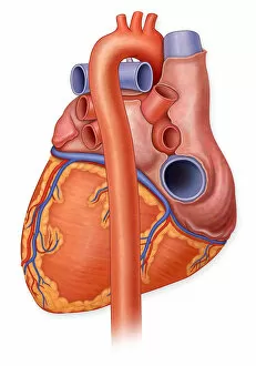
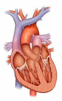
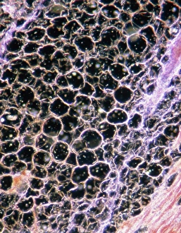
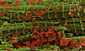
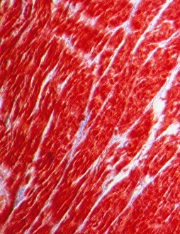
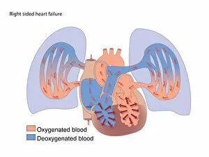
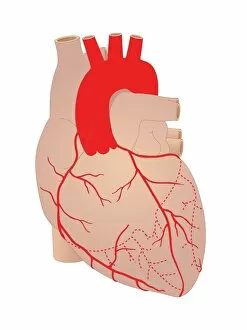
The heart muscle, also known as cardiac muscle, is a remarkable organ that plays a vital role in our body's functioning. A posterior view of a normal heart and its arteries reveals the intricate network responsible for pumping blood throughout the body. In a cross-section of this incredible muscle, we witness its complex structure and organization. Under the light microscope, we can observe the unique characteristics of cardiac muscle fibers. This microscopic view showcases both healthy tissue and fatty disease, highlighting the importance of maintaining cardiovascular health. Similarly, a colored scanning electron micrograph unveils an up-close look at vibrant and robust heart muscle fibers. Further exploration through microscopy allows us to delve into the world of striated cardiac muscles with false-color transmission electron microscopy. This technique provides valuable insights into their composition and function. However, not all conditions affecting the heart are positive. An image depicting an aortic aneurysm reminds us that certain ailments can pose serious threats to our cardiovascular system. Artwork representing right-sided and left-sided heart failure serves as poignant reminders of how crucial it is to care for our hearts diligently. Artistic renderings showcasing coronary arteries emphasize their significance in supplying oxygen-rich blood to every part of our bodies. Conversely, artwork portraying damaged cardiac tissue serves as a stark reminder that neglecting cardiovascular health can have dire consequences. Exploring various aspects related to heart muscles offers invaluable knowledge about their structure, function, diseases they may encounter like fatty disease or even more severe conditions such as right-sided or left-sided heart failure or damage due to factors like aneurysms or other complications related to coronary arteries.