
Hair Cell Collection
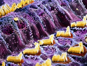
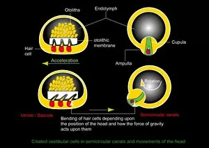
"Hair Cells: The Sensory Powerhouses of the Inner Ear" Ampullary cupula, artwork: Delicate structures that detect motion and help maintain balance in our inner ear


All Professionally Made to Order for Quick Shipping
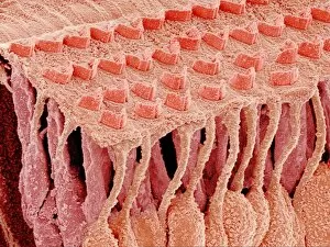
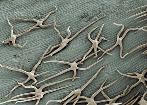
"Hair Cells: The Sensory Powerhouses of the Inner Ear" Ampullary cupula, artwork: Delicate structures that detect motion and help maintain balance in our inner ear. Structure of the cochlea, artwork: A spiral-shaped organ responsible for converting sound vibrations into electrical signals through its hair cells. Ampullary cupula, artwork: These specialized hair cells play a crucial role in detecting changes in head position and movement. Inner ear hair cells, SEM: Magnified view reveals the intricate structure of these sensory receptors that enable us to hear and perceive sound waves. Sensory hair cells in ear, SEM: Close-up image showcasing the remarkable sensitivity and complexity of these tiny hairs responsible for our auditory perception. False-color SEM of hair cells in the inner ear: Vibrant colors enhance our understanding of how different types of hair cells contribute to hearing and balance functions. Organ of Corti, SEM: This microscopic marvel within the cochlea contains rows upon rows of sensory hair cells that transform sound vibrations into electrical impulses for interpretation by our brain. Fish lateral line sense organ, artwork: Hair cell clusters found along fish's bodies allow them to sense water movements and navigate their aquatic environment effectively. Sensory hair cell in the ear, artwork: An artistic representation highlighting one single sensory receptor responsible for transmitting auditory information to our brain with astonishing precision. 10-12. Sensory hair cells in ear, SEM (repeated): Scanning electron microscopy captures stunning details as we explore further into this intricate world where countless sensory hairs work together harmoniously to provide us with an extraordinary sense - hearing.