Fraud Blocker Skip to main content

Gynaecological Collection

Gynaecological advancements have played a crucial role in women's health throughout history

Background imageGynaecological Collection: Ovarian cancer, light micrograph C015 / 7103

Ovarian cancer, light micrograph C015 / 7103
Ovary cancer. Light micrograph of a section through a Krukenberg tumour (down centre) of the ovary. This is a secondary cancer that spreads (metastasises) from the digestive system

Background imageGynaecological Collection: 885710

885710

Background imageGynaecological Collection: The first-born male offspring, the midwife, shows the newborn son to the other family members

The first-born male offspring, the midwife, shows the newborn son to the other family members
7280210 The first-born male offspring, the midwife, shows the newborn son to the other family members by Unknown Artist, (19th century); Private Collection; (add.info.: The first-born male offspring)

Background imageGynaecological Collection: Childbirth (woodcut)

Childbirth (woodcut)
7186200 Childbirth (woodcut) by Swiss School, (16th century); Private Collection; (add.info.: Childbirth. Illustration from Bilder-Lexicon Sexualwissenschaft (Verlag fur Kulturforschung)

Background imageGynaecological Collection: Woman giving birth (woodcut)

Woman giving birth (woodcut)
7201480 Woman giving birth (woodcut) by German School, (16th century) (after); Private Collection; (add.info.: Woman giving birth)

Background imageGynaecological Collection: Woman giving birth (woodcut)

Woman giving birth (woodcut)
7201479 Woman giving birth (woodcut) by German School, (16th century) (after); Private Collection; (add.info.: Woman giving birth)

Background imageGynaecological Collection: An operation on a vesicovaginal fistula, plate from Traite Complet de l Anatomie de l Homme by

An operation on a vesicovaginal fistula, plate from Traite Complet de l Anatomie de l Homme by Jean-Baptiste Marc
CHT235004 An operation on a vesicovaginal fistula, plate from Traite Complet de l Anatomie de l Homme by Jean-Baptiste Marc Bourgery (1797-1849) 1866-67 (coloured engraving) by Jacob

Background imageGynaecological Collection: Vaginal speculum used by Joseph-Claude-Anthelme Recamier (1774-1852) (wood & metal)

Vaginal speculum used by Joseph-Claude-Anthelme Recamier (1774-1852) (wood & metal)
CHT236082 Vaginal speculum used by Joseph-Claude-Anthelme Recamier (1774-1852) (wood & metal) by French School, (19th century); Musee d Histoire de la Medecine, Paris

Background imageGynaecological Collection: Female anatomy, from Commentaria cum amplissimis additionibus super anatomiam

Female anatomy, from Commentaria cum amplissimis additionibus super anatomiam
CHT236081 Female anatomy, from Commentaria cum amplissimis additionibus super anatomiam mundini, by Jacopo Berengario da Carpi (c.1460-c.1530) published in Bologna

Background imageGynaecological Collection: MS Hunter 658 Plate XXVI Drawing from William Hunters (1718-83)

MS Hunter 658 Plate XXVI Drawing from William Hunters (1718-83)
GUL232057 MS Hunter 658 Plate XXVI Drawing from William Hunters (1718-83) Anatomy of the Human Gravid Uterus, 1774 (chalk on paper) by Rymsdyk, Jan van (fl.1750-88/89); Glasgow University Library

Background imageGynaecological Collection: Forceps used by Jan Palfyn of Ghent (1650-1730) (wood & metal)

Forceps used by Jan Palfyn of Ghent (1650-1730) (wood & metal)
CHT236080 Forceps used by Jan Palfyn of Ghent (1650-1730) (wood & metal) by French School, (18th century); Musee d Histoire de la Medecine, Paris, France; eArchives Charmet; French, out of copyright

Background imageGynaecological Collection: A night of fire in La Spezia (colour litho)

A night of fire in La Spezia (colour litho)
2784248 A night of fire in La Spezia (colour litho) by Beltrame, Achille (1871-1945); Private Collection; (add.info.: A young midwife is called urgently and rushes to a house that is ruining)

Background imageGynaecological Collection: Vaginal speculum used by Antoine Jobert de Lamballe (1799-1867) (metal & ivory)

Vaginal speculum used by Antoine Jobert de Lamballe (1799-1867) (metal & ivory)
CHT236088 Vaginal speculum used by Antoine Jobert de Lamballe (1799-1867) (metal & ivory) by French School, (19th century); Musee d Histoire de la Medecine, Paris, France; eArchives Charmet; French

Background imageGynaecological Collection: Anatomy of a pregnant woman, from Manuel d Anatomie descriptive du Corps

Anatomy of a pregnant woman, from Manuel d Anatomie descriptive du Corps
CHT236084 Anatomy of a pregnant woman, from Manuel d Anatomie descriptive du Corps Humain by Jules Cloquet (1790-1883) engraved by G

Background imageGynaecological Collection: Midwifery, illustration from Encyclopedia Britannica, published in Edinburgh

Midwifery, illustration from Encyclopedia Britannica, published in Edinburgh
DGC398035 Midwifery, illustration from Encyclopedia Britannica, published in Edinburgh, 1798 (copperplate engraving) by Bell, Alexander (fl)

Background imageGynaecological Collection: Statuette of a woman giving birth, given to pregnant women for a successful delivery

Statuette of a woman giving birth, given to pregnant women for a successful delivery
CHT236071 Statuette of a woman giving birth, given to pregnant women for a successful delivery, from Alexandria (terracotta) by Egyptian Ptolemaic Period (332-30 BC); Private Collection; (add.info)

Background imageGynaecological Collection: Ovarian cyst, endoscope view C017 / 6800

Ovarian cyst, endoscope view C017 / 6800
Ovarian cyst. Endoscope (laparascope) view of a cyst (white) on a patients ovary. Ovarian cysts are common in women of child-bearing age

Background imageGynaecological Collection: Uterus, endoscope view C017 / 6805

Uterus, endoscope view C017 / 6805
Uterus. Endoscope (laparascope) view of a healthy uterus (womb, large, upper centre), with the two fallopian tubes (left and right) and an ovary (white, lower right)

Background imageGynaecological Collection: Menopause, conceptual artwork

Menopause, conceptual artwork. Menopause is the cessation of menstruation. It usually occurs between the ages of 45 and 55

Background imageGynaecological Collection: Heavy menstrual bleeding, artwork

Heavy menstrual bleeding, artwork
Heavy menstrual bleeding, conceptual computer artwork. Collection of measuring cylinders containing differing amounts of blood

Background imageGynaecological Collection: A Gynaecological Operation in St Petersburg

A Gynaecological Operation in St Petersburg
Early 20th century Gynaecological surgery taking place in front of a vast audience of Surgeons, would-be surgeons and nurses in the Obstetric Institute in St Petersburg, Russia. Date: 1905

Background imageGynaecological Collection: Ovarian cyst, endoscope view C017 / 6801

Ovarian cyst, endoscope view C017 / 6801
Ovarian cyst. Endoscope (laparascope) view of a cyst (white) on a patients ovary. Ovarian cysts are common in women of child-bearing age

Background imageGynaecological Collection: Ovary, endoscope view C017 / 6803

Ovary, endoscope view C017 / 6803
Ovary. Endoscope (laparascope) view of a healthy ovary (white). There are two ovaries in the female body, which release an egg (ovum) during the monthly ovulation cycle

Background imageGynaecological Collection: Ovarian cyst, endoscope view C017 / 6802

Ovarian cyst, endoscope view C017 / 6802
Ovarian cyst. Endoscope (laparascope) view of a cyst (white) on a patients ovary. Ovarian cysts are common in women of child-bearing age

Background imageGynaecological Collection: Ectopic pregnancy, endoscope view C017 / 6804

Ectopic pregnancy, endoscope view C017 / 6804
Ectopic pregnancy. Endoscope (laparascope) view of an ectopic pregnancy in a patients fallopian tube. An ectopic pregnancy (eccysis)

Background imageGynaecological Collection: IVF treatment, light microscope C016 / 6536

IVF treatment, light microscope C016 / 6536
IVF treatment. Light microscope of human egg cells (large, round) and sperm (small dark ovals with tails) in a petri dish during in vitro fertilisation (IVF)

Background imageGynaecological Collection: IVF treatment, light microscope C016 / 6535

IVF treatment, light microscope C016 / 6535
IVF treatment. Light microscope of human egg cells (large, round) and sperm (small dark ovals with tails) in a petri dish during in vitro fertilisation (IVF)

Background imageGynaecological Collection: IVF treatment, light microscope C016 / 6512

IVF treatment, light microscope C016 / 6512
IVF treatment. Light microscope of human egg cells (large, round) and sperm (small dark ovals with tails) in a petri dish during in vitro fertilisation (IVF)

Background imageGynaecological Collection: Vaginal douche, circa 1900 C018 / 0357

Vaginal douche, circa 1900 C018 / 0357
Vaginal douche by Dr. Eguisier. Douches such as this one, although unreliable, were one of the most common methods of contraception in this era

Background imageGynaecological Collection: Uterine fibroid, X-ray C014 / 4919

Uterine fibroid, X-ray C014 / 4919
Uterus tumour. Coloured X-ray of the abdomen of a woman with a fibroid (round, centre) in her uterus. A fibroid is a fibrous benign tumour originating from muscular tissue (leimyoma)

Background imageGynaecological Collection: Uterine fibroid, X-ray C014 / 4918

Uterine fibroid, X-ray C014 / 4918
Uterus tumour. Coloured X-ray of the abdomen of a woman with a fibroid (orange, round) in her uterus. A fibroid is a fibrous benign tumour originating from muscular tissue (leimyoma)

Background imageGynaecological Collection: Endometriosis, X-ray C014 / 4917

Endometriosis, X-ray C014 / 4917
Endometriosis. Coloured X-ray of the abdomen of woman with endometriosis. Endometriosis is the movement of cells from the lining of the uterus to the abdominal cavity

Background imageGynaecological Collection: Endometriosis, X-ray C014 / 4916

Endometriosis, X-ray C014 / 4916
Endometriosis. Coloured X-ray of the abdomen of woman with endometriosis. Endometriosis is the movement of cells from the lining of the uterus to the abdominal cavity

Background imageGynaecological Collection: Dermoid ovarian cyst C015 / 6910

Dermoid ovarian cyst C015 / 6910
Dermoid ovarian cyst. Light micrograph of a section through an ovary with a dermoid, or teratoma, ovarian cyst. This is a benign (non-cancerous) cyst that arises from germ (sex) cells

Background imageGynaecological Collection: Cancer of the chorion C015 / 6905

Cancer of the chorion C015 / 6905
Cancer of the chorion. Light micrograph of a section through a sarcoma of the chorion, a malignant (cancerous) tumour of the membrane (chorion) that surrounds a developing embryo

Background imageGynaecological Collection: Endometrial polyp C015 / 6745

Endometrial polyp C015 / 6745
Endometrial polyp. Gross clinical specimen of a polyp (centre) in the uterus (womb). This polyp, a benign (non-cancerous) growth, has arisen from the endometrium, the lining of the uterus

Background imageGynaecological Collection: Endometrial polyp C015 / 6742

Endometrial polyp C015 / 6742
Endometrial polyp. Gross clinical specimen of a polyp (centre) in the uterus (womb). This polyp, a benign (non-cancerous) growth, has arisen from the endometrium, the lining of the uterus

Background imageGynaecological Collection: Dermoid ovarian cyst C015 / 6056

Dermoid ovarian cyst C015 / 6056
Dermoid ovarian cyst. Light micrograph of a section through an ovary with a dermoid, or teratoma, ovarian cyst. This is a benign (non-cancerous) cyst that arises from germ (sex) cells

Background imageGynaecological Collection: Cervical thickening, light micrograph C015 / 6166

Cervical thickening, light micrograph C015 / 6166
Cervical thickening. Light micrograph of a section through the thickened mucous membrane of a cervix (neck of the womb). This condition, known as leukoplakia, occurs in 1% of elderly women

Background imageGynaecological Collection: Dermoid ovarian cyst C015 / 6402

Dermoid ovarian cyst C015 / 6402
Dermoid ovarian cyst. Light micrograph of a section through an ovary with a dermoid, or teratoma, ovarian cyst. This is a benign (non-cancerous) cyst that arises from germ (sex) cells

Background imageGynaecological Collection: Cervical thickening, light micrograph C015 / 6167

Cervical thickening, light micrograph C015 / 6167
Cervical thickening. Light micrograph of a section through the thickened mucous membrane (dark purple, left) of a cervix (neck of the womb). Normal epithelium is at right

Background imageGynaecological Collection: Cervical thickening, light micrograph C015 / 6168

Cervical thickening, light micrograph C015 / 6168
Cervical thickening. Light micrograph of a section through the thickened mucous membrane (dark purple, right) of a cervix (neck of the womb). Normal epithelium is at left

Background imageGynaecological Collection: Artwork of retroflexion of the uterus

Artwork of retroflexion of the uterus

Background imageGynaecological Collection: Blocked Fallopian tubes, X-ray

Blocked Fallopian tubes, X-ray
Blocked Fallopian tubes. Coloured X-ray of a womans reproductive system, with her uterus (green) and blocked Fallopian tubes (oviducts, yellow)

Background imageGynaecological Collection: Heavy menstrual bleeding

Heavy menstrual bleeding. Conceptual computer artwork of red blood cells pouring out a tap, representing the heavy bleeding seen in menorrhagia, or excessive menstrual bleeding

Background imageGynaecological Collection: Endometriosis

Endometriosis. Computer artwork of endrometriomas, masses of cells from the lining of the uterus, thickening (right) and bleeding (bottom)

Background imageGynaecological Collection: Uterine fibroid, MRI scan

Uterine fibroid, MRI scan

Background imageGynaecological Collection: Endometriosis, computer artwork

Endometriosis, computer artwork
Endometriosis. Conceptual computer artwork of endometriosis, the movement of cells from the lining of the uterus to the abdominal cavity


All Professionally Made to Order for Quick Shipping

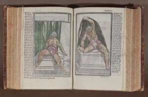
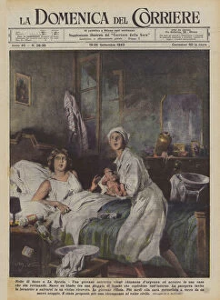
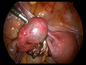
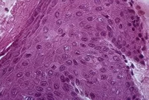
Gynaecological advancements have played a crucial role in women's health throughout history. From the early understanding of female anatomy to modern medical interventions, these hints shed light on the progress made in this field. Ovarian cancer, a devastating disease affecting countless women worldwide, has been extensively studied by researchers and healthcare professionals. The light micrograph C015/7103 provides valuable insights into the cellular structure of this malignant condition, aiding in its diagnosis and treatment. In ancient times, childbirth was surrounded by mystery and often left to midwives who possessed knowledge passed down through generations. The heartwarming image of a midwife proudly presenting the first-born male offspring to other family members showcases the importance placed on new life and highlights the pivotal role midwives played in ensuring safe deliveries. The meticulous drawings by Ms Hunter for William Hunter's Anatomy of the Human Gravid Uterus offer an artistic representation of pregnancy during the 18th century. These detailed chalk sketches provide a glimpse into our ancestors' understanding of fetal development within the womb, contributing significantly to obstetric knowledge at that time. Examining female genitals from painted cardboard dating back to approximately 1830 reveals how anatomical studies evolved over centuries. Such visual aids were instrumental in educating medical students about reproductive structures and their functions. Woodcut illustrations depicting childbirth remind us of historical practices surrounding labor and delivery. These images capture both the pain endured by women during this natural process as well as society's fascination with witnessing such moments firsthand. Not all gynaecological conditions are related to pregnancy; some require surgical intervention for proper treatment. An engraving from Traite Complet de l'Anatomie de l'Homme depicts an operation performed on a vesicovaginal fistula—an abnormal connection between bladder and vagina—highlighting surgical techniques developed over time to address complex gynaecological issues successfully.