Fraud Blocker Skip to main content

Grays Anatomy Collection

"Exploring the Intricacies of Anatomy

Background imageGrays Anatomy Collection: Bones of the left hand, Dorsal surface (engraving)

Bones of the left hand, Dorsal surface (engraving)
3634211 Bones of the left hand, Dorsal surface (engraving) by English School, (19th century); Private Collection; (add.info.: Bones of the left hand, Dorsal surface)

Background imageGrays Anatomy Collection: Bones of the right foot, Plantar surface (engraving)

Bones of the right foot, Plantar surface (engraving)
3634207 Bones of the right foot, Plantar surface (engraving) by English School, (19th century); Private Collection; (add.info.: Bones of the right foot, Plantar surface)

Background imageGrays Anatomy Collection: Left hip-joint laid open (engraving)

Left hip-joint laid open (engraving)
3634205 Left hip-joint laid open (engraving) by English School, (19th century); Private Collection; (add.info.: Left hip-joint laid open)

Background imageGrays Anatomy Collection: Muscles of the tongue from below (engraving)

Muscles of the tongue from below (engraving)
3634201 Muscles of the tongue from below (engraving) by English School, (19th century); Private Collection; (add.info.: Muscles of the tongue from below)

Background imageGrays Anatomy Collection: The abdominal aorta and its branches (engraving)

The abdominal aorta and its branches (engraving)
3634189 The abdominal aorta and its branches (engraving) by English School, (19th century); Private Collection; (add.info.: The abdominal aorta and its branches)

Background imageGrays Anatomy Collection: Upper surface of the tongue (engraving)

Upper surface of the tongue (engraving)
3634180 Upper surface of the tongue (engraving) by English School, (19th century); Private Collection; (add.info.: Upper surface of the tongue)

Background imageGrays Anatomy Collection: The permanent teeth, external view (engraving)

The permanent teeth, external view (engraving)
3634176 The permanent teeth, external view (engraving) by English School, (19th century); Private Collection; (add.info.: The permanent teeth, external view)

Background imageGrays Anatomy Collection: The roots of the lungs and posterior pulmonary plexus, seen from behind (engraving)

The roots of the lungs and posterior pulmonary plexus, seen from behind (engraving)
3634173 The roots of the lungs and posterior pulmonary plexus, seen from behind (engraving) by English School, (19th century); Private Collection; (add.info)

Background imageGrays Anatomy Collection: Muscles of the left hand, Palmar surface (engraving)

Muscles of the left hand, Palmar surface (engraving)
3634195 Muscles of the left hand, Palmar surface (engraving) by English School, (19th century); Private Collection; (add.info.: Muscles of the left hand, Palmar surface)

Background imageGrays Anatomy Collection: The sympathetic nerve (engraving)

The sympathetic nerve (engraving)
3634182 The sympathetic nerve (engraving) by English School, (19th century); Private Collection; (add.info.: The sympathetic nerve)

Background imageGrays Anatomy Collection: A front view of the organ of hearing, right side (engraving)

A front view of the organ of hearing, right side (engraving)
3634179 A front view of the organ of hearing, right side (engraving) by English School, (19th century); Private Collection; (add.info.: A front view of the organ of hearing, right side)

Background imageGrays Anatomy Collection: The pancreas and its relations (engraving)

The pancreas and its relations (engraving)
3634174 The pancreas and its relations (engraving) by English School, (19th century); Private Collection; (add.info.: The pancreas and its relations)

Background imageGrays Anatomy Collection: Frontal bone, outer surface (engraving)

Frontal bone, outer surface (engraving)
3634209 Frontal bone, outer surface (engraving) by English School, (19th century); Private Collection; (add.info.: Frontal bone, outer surface)

Background imageGrays Anatomy Collection: Muscles on the dorsum of the tongue (engraving)

Muscles on the dorsum of the tongue (engraving)
3634200 Muscles on the dorsum of the tongue (engraving) by English School, (19th century); Private Collection; (add.info.: Muscles on the dorsum of the tongue)

Background imageGrays Anatomy Collection: The arch of the aorta and its branches (engraving)

The arch of the aorta and its branches (engraving)
3634194 The arch of the aorta and its branches (engraving) by English School, (19th century); Private Collection; (add.info.: The arch of the aorta and its branches)

Background imageGrays Anatomy Collection: The internal carotid and vertbral arteries, right side (engraving)

The internal carotid and vertbral arteries, right side (engraving)
3634190 The internal carotid and vertbral arteries, right side (engraving) by English School, (19th century); Private Collection; (add.info.: The internal carotid and vertbral arteries, right side)

Background imageGrays Anatomy Collection: Gray matter of the cerebral cortex, Meynert (engraving)

Gray matter of the cerebral cortex, Meynert (engraving)
3634183 Gray matter of the cerebral cortex, Meynert (engraving) by English School, (19th century); Private Collection; (add.info.: Gray matter of the cerebral cortex, Meynert)

Background imageGrays Anatomy Collection: Palmar fascia (engraving)

Palmar fascia (engraving)
3634196 Palmar fascia (engraving) by English School, (19th century); Private Collection; (add.info.: Palmar fascia. Illustration for Grays Anatomy, ie Anatomy, Descriptive and Surgical by Henry Gray)

Background imageGrays Anatomy Collection: Occipital bone, inner surface (engraving)

Occipital bone, inner surface (engraving)
3634210 Occipital bone, inner surface (engraving) by English School, (19th century); Private Collection; (add.info.: Occipital bone, inner surface)

Background imageGrays Anatomy Collection: Bones of the right foot, Dorsal surface (engraving)

Bones of the right foot, Dorsal surface (engraving)
3634206 Bones of the right foot, Dorsal surface (engraving) by English School, (19th century); Private Collection; (add.info.: Bones of the right foot, Dorsal surface)

Background imageGrays Anatomy Collection: The arteries of the face and scalp (engraving)

The arteries of the face and scalp (engraving)
3634192 The arteries of the face and scalp (engraving) by English School, (19th century); Private Collection; (add.info.: The arteries of the face and scalp)

Background imageGrays Anatomy Collection: Drawing to illustrate cranio-cerebral topography, Macalister (engraving)

Drawing to illustrate cranio-cerebral topography, Macalister (engraving)
3634186 Drawing to illustrate cranio-cerebral topography, Macalister (engraving) by English School, (19th century); Private Collection; (add.info.: Drawing to illustrate cranio-cerebral topography)

Background imageGrays Anatomy Collection: Cartilages of the nose (engraving)

Cartilages of the nose (engraving)
3634181 Cartilages of the nose (engraving) by English School, (19th century); Private Collection; (add.info.: Cartilages of the nose)

Background imageGrays Anatomy Collection: The Diaphragm, under surface (engraving)

The Diaphragm, under surface (engraving)
3634199 The Diaphragm, under surface (engraving) by English School, (19th century); Private Collection; (add.info.: The Diaphragm, under surface)

Background imageGrays Anatomy Collection: Muscles of the chest and front of the arm, superficial view (engraving)

Muscles of the chest and front of the arm, superficial view (engraving)
3634197 Muscles of the chest and front of the arm, superficial view (engraving) by English School, (19th century); Private Collection; (add.info.: Muscles of the chest and front of the arm)

Background imageGrays Anatomy Collection: Topography of thoracic and abdominal viscera (engraving)

Topography of thoracic and abdominal viscera (engraving)
3634177 Topography of thoracic and abdominal viscera (engraving) by English School, (19th century); Private Collection; (add.info.: Topography of thoracic and abdominal viscera)

Background imageGrays Anatomy Collection: The salivary glands (engraving)

The salivary glands (engraving)
3634175 The salivary glands (engraving) by English School, (19th century); Private Collection; (add.info.: The salivary glands)

Background imageGrays Anatomy Collection: Muscles of the head, face, and neck (engraving)

Muscles of the head, face, and neck (engraving)
3634202 Muscles of the head, face, and neck (engraving) by English School, (19th century); Private Collection; (add.info.: Muscles of the head, face, and neck)

Background imageGrays Anatomy Collection: Side view of pelvis, showing Levator ani (engraving)

Side view of pelvis, showing Levator ani (engraving)
3634198 Side view of pelvis, showing Levator ani (engraving) by English School, (19th century); Private Collection; (add.info.: Side view of pelvis, showing Levator ani)

Background imageGrays Anatomy Collection: The arteries of the base of the brain (engraving)

The arteries of the base of the brain (engraving)
3634191 The arteries of the base of the brain (engraving) by English School, (19th century); Private Collection; (add.info.: The arteries of the base of the brain)

Background imageGrays Anatomy Collection: Veins of the head and neck (engraving)

Veins of the head and neck (engraving)
3634188 Veins of the head and neck (engraving) by English School, (19th century); Private Collection; (add.info.: Veins of the head and neck)

Background imageGrays Anatomy Collection: Magnified view of the human embryo of four weeks, with the membranes opened (engraving)

Magnified view of the human embryo of four weeks, with the membranes opened (engraving)
3634214 Magnified view of the human embryo of four weeks, with the membranes opened (engraving) by English School, (19th century); Private Collection; (add.info)

Background imageGrays Anatomy Collection: Occipital bone, outer surface (engraving)

Occipital bone, outer surface (engraving)
3634213 Occipital bone, outer surface (engraving) by English School, (19th century); Private Collection; (add.info.: Occipital bone, outer surface)

Background imageGrays Anatomy Collection: Bones of the left hand, Palmar surface (engraving)

Bones of the left hand, Palmar surface (engraving)
3634208 Bones of the left hand, Palmar surface (engraving) by English School, (19th century); Private Collection; (add.info.: Bones of the left hand, Palmar surface)

Background imageGrays Anatomy Collection: Longitudinal section through the middle of the right knee-joint (engraving)

Longitudinal section through the middle of the right knee-joint (engraving)
3634204 Longitudinal section through the middle of the right knee-joint (engraving) by English School, (19th century); Private Collection; (add.info)

Background imageGrays Anatomy Collection: Surgical anatomy of the arteries of the neck, right side (engraving)

Surgical anatomy of the arteries of the neck, right side (engraving)
3634193 Surgical anatomy of the arteries of the neck, right side (engraving) by English School, (19th century); Private Collection; (add.info.: Surgical anatomy of the arteries of the neck)

Background imageGrays Anatomy Collection: The superficial lymphatics and glands of the head, face, and neck (engraving)

The superficial lymphatics and glands of the head, face, and neck (engraving)
3634187 The superficial lymphatics and glands of the head, face, and neck (engraving) by English School, (19th century); Private Collection; (add.info)

Background imageGrays Anatomy Collection: The deep lymphatics and glands of the neck and thorax (engraving)

The deep lymphatics and glands of the neck and thorax (engraving)
3634185 The deep lymphatics and glands of the neck and thorax (engraving) by English School, (19th century); Private Collection; (add.info.: The deep lymphatics and glands of the neck and thorax)

Background imageGrays Anatomy Collection: Base of the brain (engraving)

Base of the brain (engraving)
3634184 Base of the brain (engraving) by English School, (19th century); Private Collection; (add.info.: Base of the brain)

Background imageGrays Anatomy Collection: Face of an embryo of 25 to 28 days, magnified 15 times (engraving)

Face of an embryo of 25 to 28 days, magnified 15 times (engraving)
3634212 Face of an embryo of 25 to 28 days, magnified 15 times (engraving) by English School, (19th century); Private Collection; (add.info.: Face of an embryo of 25 to 28 days, magnified 15 times)

Background imageGrays Anatomy Collection: Title page of Henry Grays Anatomy, 1858

Title page of Henry Grays Anatomy, 1858
GRAYs ANATOMY, 1858. Title page of Henry Grays Anatomy, 1858


All Professionally Made to Order for Quick Shipping

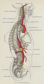
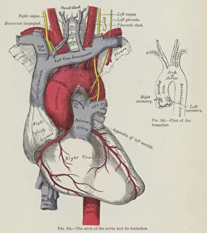
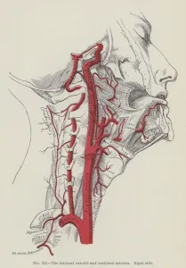
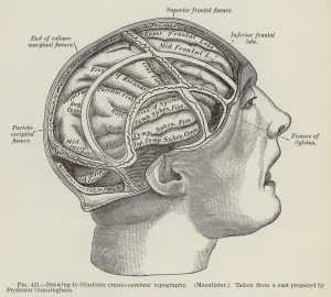
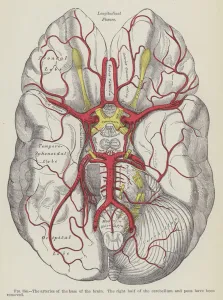
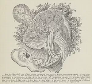
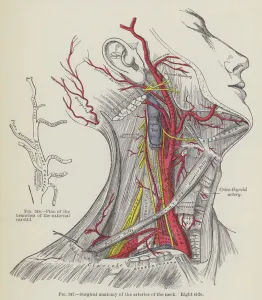
"Exploring the Intricacies of Anatomy: A Journey through Gray's Anatomy Engravings" Step into the world of anatomy as we delve into the mesmerizing engravings from Gray's Anatomy. From the intricate bones of the left hand, meticulously depicted on its dorsal surface, to a captivating portrayal of the bones on the right foot's plantar surface – each engraving unveils a hidden realm within our bodies. Witness an astonishing revelation as you explore a laid-open left hip-joint, exposing its inner workings and shedding light on its complex structure. Marvel at the muscles of the tongue captured from below, showcasing their remarkable intricacy and functionality. Embark on an educational voyage through engravings that bring to life our internal systems. Observe with awe as we unravel the mysteries surrounding abdominal aorta and its branches – vital lifelines coursing through our bodies. Delve deeper into oral health as you examine an enchanting depiction of upper surface of tongue alongside external view illustrations capturing permanent teeth in all their glory. Gaze upon breathtaking renditions unveiling roots of lungs and posterior pulmonary plexus seen from behind – offering us insights into respiratory marvels concealed beneath our skin. Immerse yourself in understanding every muscle that adorns your hand's palmar surface, appreciating their dexterity and strength. Discover how nerves play a crucial role in orchestrating bodily functions by exploring detailed depictions illustrating sympathetic nerve pathways. Witness an organ like no other come alive before your eyes - experience a front view engraving revealing intricate details about hearing mechanisms found within. Lastly, be captivated by engravings showcasing pancreas and its relationships with neighboring organs - unlocking secrets about this essential gland responsible for regulating digestion. Gray's Anatomy engravings offer us glimpses into human complexity beyond what meets our eyes. Join us on this enlightening journey where artistry meets science, unraveling wonders hidden beneath our skin.