
Granulocytes Collection
Granulocytes are a crucial component of our immune system, playing a vital role in defending our bodies against infections and diseases


All Professionally Made to Order for Quick Shipping
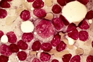
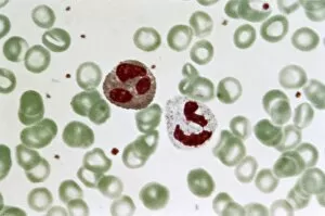
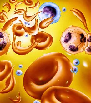
Granulocytes are a crucial component of our immune system, playing a vital role in defending our bodies against infections and diseases. These specialized white blood cells can be found in various forms, each with its unique characteristics. One such condition where they can affected is chronic lymphocytic leukemia (CLL). In the micrograph of CLL, we can observe abnormal granulocyte development, which hampers their ability to fight off infections effectively. Another disorder that impacts granulocytes is Chediak-Higashi syndrome. The micrograph reveals enlarged granules within these cells, leading to impaired functioning and increased susceptibility to infections. Chronic myeloid leukemia (CML) also affects granulocytes. Micrographs of CML display an excessive number of immature granulocytes called blasts, indicating uncontrolled growth and potential complications. In contrast, a light micrograph showcases healthy granulocyte blood cells at work. These powerful defenders patrol our bloodstream ready to engulf any foreign invaders they encounter. Moving back to CML but from another perspective - the micrograph C015/6227 highlights the presence of blast crisis cells in this type of leukemia. This image emphasizes the urgency for timely diagnosis and treatment to prevent further progression. Acute myeloid leukemia (AML), as seen in micrograph C015/6225, displays abnormal maturation patterns within granulocyte precursors. This disruption compromises their functionality and weakens the body's defense mechanisms against pathogens. Apart from their role in immunity, not all microscopic images revolve around diseases; some focus on other cell types like fat cells observed through scanning electron microscopy (SEM). These images provide insights into adipose tissue structure and function while reminding us that there's more than one type of cell worth studying under a microscope. However, it's important not to overlook conditions like sickle cell anemia or psoriasis when discussing human blood cells' diversity.