Fraud Blocker Skip to main content

Glial Cells Collection

Glial cells, also known as neuroglia or glia, are an essential component of the nervous system

Background imageGlial Cells Collection: Brain cancer, DTI and 3D CT scans C016 / 6414

Brain cancer, DTI and 3D CT scans C016 / 6414
Brain cancer. Coloured composite image of diffusion tensor imaging (DTI) scans of the brains, and 3D computed tomography (CT) scans of the skulls, of two 35 year old patients

Background imageGlial Cells Collection: Schematic of the hypothalamus receiving nerve impulses from the body

Schematic of the hypothalamus receiving nerve impulses from the body and sending messages to the circulatory and nervous system

Background imageGlial Cells Collection: Nerve with myelin sheath, seen in lower right, connects with muscle

Nerve with myelin sheath, seen in lower right, connects with muscle. Blood vessel and immune cells are seen in the center and upper right of image

Background imageGlial Cells Collection: Neurofibromatosis, a genetic disorder of the nervous system

Neurofibromatosis, a genetic disorder of the nervous system
A genetic disorder of the nervous system, neurofibromatosis causes tumors to form on nerves throughout the body, including a type of tumor called an optic nerve glioma that can result in childhood

Background imageGlial Cells Collection: Directed differentiation of multipotential human neural progenitor cells

Directed differentiation of multipotential human neural progenitor cells
Human neural progenitor cells were isolated under selective culture conditions from the developing human brain and directed through lineage differentiation to GFAP + (glial fibrillary acid protein)

Background imageGlial Cells Collection: Nerve ganglion, light micrograph C016 / 0532

Nerve ganglion, light micrograph C016 / 0532
Nerve ganglion. Light micrograph of a section through a nerve ganglion of the peripheral nervous system, showing clusters of nerve cell bodies

Background imageGlial Cells Collection: Brain cancer, MRI scan C016 / 4438

Brain cancer, MRI scan C016 / 4438
Brain cancer. Coloured coronal magnetic resonance imaging (MRI) scan through the head of a 48 year old male patient with a glioblastoma (red)

Background imageGlial Cells Collection: Brain cancer, MRI scan C016 / 4436

Brain cancer, MRI scan C016 / 4436
Brain cancer. Coloured coronal magnetic resonance imaging (MRI) scan through the head of a 48 year old male patient with a glioblastoma (centre right)

Background imageGlial Cells Collection: Brain cancer, MRI scan C016 / 4437

Brain cancer, MRI scan C016 / 4437
Brain cancer. Coloured coronal magnetic resonance imaging (MRI) scan through the head of a 48 year old male patient with a glioblastoma (orange)

Background imageGlial Cells Collection: Spinal nerve ganglion, light micrograph

Spinal nerve ganglion, light micrograph
Spinal nerve ganglion. Light micrograph of a cross-section through a spinal nerve ganglion. This is a node of nerve cells located just outside the spinal cord at the point where it is joined by

Background imageGlial Cells Collection: Brain lining, SEM

Brain lining, SEM
Brain lining. Coloured scanning electron micrograph (SEM) of the lining of the brain, showing the ciliary hairs (green) of ependymal cells


All Professionally Made to Order for Quick Shipping

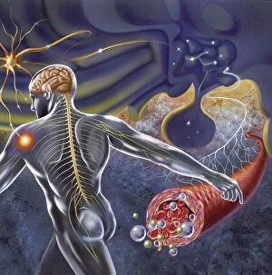
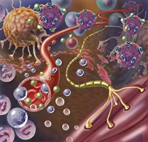
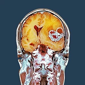
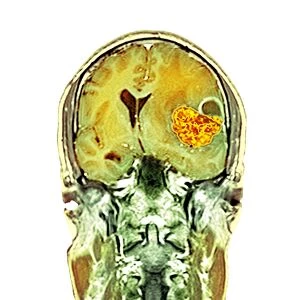
Glial cells, also known as neuroglia or glia, are an essential component of the nervous system. These remarkable cells play a crucial role in supporting and protecting neurons, the primary functional units of the brain. When we examine the brain lining under a scanning electron microscope (SEM), we can observe intricate networks intertwined with neurons. They form a complex web that provides structural support and insulation to these delicate nerve cells. In another SEM image, we see glial cells present in Fallopian tube tissue. Although their function here is not fully understood yet, it highlights how versatile these cells are throughout different parts of our body. However, sometimes things go awry within our neural network. Brain cancer becomes apparent through advanced imaging techniques such as diffusion tensor imaging (DTI) and 3D CT scans. These scans reveal abnormal growths within the brain's delicate structures – a stark reminder of how vital glial cell functioning is for maintaining neurological health. A schematic representation showcases how the hypothalamus receives nerve impulses from various parts of our body. Glial cells surround these connections, ensuring efficient communication between different regions and coordinating bodily functions seamlessly. Moving down to microscopic levels, we encounter nerves with myelin sheaths connecting with muscles – an illustration of how glial cells aid in transmitting signals effectively across long distances within our bodies. Unfortunately, genetic disorders like neurofibromatosis disrupt this harmony. This condition affects the nervous system and often leads to tumors forming on nerves themselves or nearby tissues – highlighting once again why understanding glial cell behavior is critical for combating such diseases. Scientists have made significant strides in directed differentiation experiments involving multipotential human neural progenitor cells – unlocking potential therapeutic avenues by manipulating these versatile glial precursors into specific neuronal lineages. Examining light micrographs reveals fascinating details about nerve ganglia where clusters of neuronal cell bodies reside alongside supportive glia - further emphasizing their indispensable role in maintaining neural function.