Fraud Blocker Skip to main content

Glandular Collection (page 2)

"Glandular: Unveiling the Intricacies of Nature's Secretive World" Botanik Digitalis purpurea L

Background imageGlandular Collection: Round-leaved Sundew (Drosera rotundifolia) glandular hairs on leaf with sticky mucilage, Italy, june

Round-leaved Sundew (Drosera rotundifolia) glandular hairs on leaf with sticky mucilage, Italy, june

Background imageGlandular Collection: Fly glandular organ, SEM

Fly glandular organ, SEM
Fly glandular organ. Coloured scanning electron micrograph (SEM) of a glandular organ on a March fly (family Bibionidae). This organ is found on the rear of the fly

Background imageGlandular Collection: Hair follicles, SEM

Hair follicles, SEM
Hair follicles. Coloured scanning electron micrograph (SEM) of a section through freeze- fractured hair follicles in the skin

Background imageGlandular Collection: Pituitary gland, TEM

Pituitary gland, TEM
Pituitary gland. Coloured transmission electron micrograph (TEM) of cells in the anterior pituitary gland, a hormone-secreting gland at the base of the brain

Background imageGlandular Collection: Secretory cells in pancreas, SEM

Secretory cells in pancreas, SEM
Pancreatic secretory cells. Coloured scanning electron micrograph (SEM) of a freeze-fracture through a healthy pancreas, showing the secretory tissue

Background imageGlandular Collection: Lactating breast tissue, SEM

Lactating breast tissue, SEM

Background imageGlandular Collection: Lacrimal gland, SEM

Lacrimal gland, SEM

Background imageGlandular Collection: Blood vessels from a salivary gland, SEM

Blood vessels from a salivary gland, SEM
Blood vessels from a submandibular (submaxillary) salivary gland, coloured scanning electron micrograph (SEM). This is a cast: the vessels were filled with a resin, which then set

Background imageGlandular Collection: Breast implant calcification, X-ray

Breast implant calcification, X-ray
Breast implant calcification. Coloured mammogram (breast X-ray) of calcification around the borders of a breast implant (pink) in a womans breast (red) as seen from the side

Background imageGlandular Collection: Ulcerative colitis, SEM

Ulcerative colitis, SEM
Ulcerative colitis. Coloured scanning electron micrograph (SEM) of freeze-fractured bowel tissue from a patient suffering from ulcerative colitis

Background imageGlandular Collection: Stomach lining with gastric pits, SEM

Stomach lining with gastric pits, SEM
Stomach lining. Coloured scanning electron micrograph (SEM) of the inner lining of the stomach (gastric mucosa). The large folds are known as rugae

Background imageGlandular Collection: Trachea mucous membrane, SEM

Trachea mucous membrane, SEM
Trachea mucous membrane. Coloured scanning electron micrograph (SEM) of a fractured mucous membrane of the trachea (wind pipe), showing the epithelium and underlying connective tissue

Background imageGlandular Collection: Marjoram leaf surface, SEM

Marjoram leaf surface, SEM
Marjoram (Origanum vulgare) leaf surface, coloured scanning electron micrograph (SEM). Glandular trichomes are yellow, hair-like trichomes are blue

Background imageGlandular Collection: Mint leaf surface, SEM

Mint leaf surface, SEM
Mint leaf surface. Coloured scanning electron micrograph (SEM) of the surface of a mint (Menta sp.) leaf. The white structures are oil glands

Background imageGlandular Collection: Intestinal villi anatomy, artwork

Intestinal villi anatomy, artwork
Intestinal villi anatomy. Artwork showing six types of cell found on the surface of villi in the small intestine. Clockwise from upper right they are: enterocytes (green)

Background imageGlandular Collection: Exocrine gland types, artwork

Exocrine gland types, artwork
Exocrine gland types. Computer artwork of the six forms an exocrine gland can take. Clockwise from bottom left these are; simple acinar, simple tubular, simple coiled tubular, simple branched acinar

Background imageGlandular Collection: Exocrine gland, artwork

Exocrine gland, artwork
Exocrine gland. Computer artwork of a section through an exocrine gland. Exocrine glands secrete their products through ducts on to epithelial surfaces

Background imageGlandular Collection: Hairs and glands on thyme

Hairs and glands on thyme
False-colour scanning electron micrograph of the leaf surface of the herb thyme, Thymus vulgaris. The micrograph shows two types of trichomes (projections from the leaf surface)

Background imageGlandular Collection: Grape vine, SEM

Grape vine, SEM
Grape vine. Coloured scanning electron micrograph (SEM) of the surface of a grape vine (Vitis sp.). It is covered in hairs and glandular trichomes (orange)

Background imageGlandular Collection: Anther gland cell, SEM

Anther gland cell, SEM
Anther gland cell. Coloured scanning electron micrograph (SEM) of a gland cell on the surface of an anther. Gland cells secrete oils and other substances to keep a plant healthy

Background imageGlandular Collection: Anther gland cells, SEM

Anther gland cells, SEM
Anther gland cells. Coloured scanning electron micrograph (SEM) of two gland cells on the surface of an anther. Gland cells secrete oils and other substances to keep a plant healthy

Background imageGlandular Collection: Rothrocks nama (Nama rothrockii)

Rothrocks nama (Nama rothrockii)
Rothrocks nama flowers (Nama rothrockii). Photographed at around 2400 metres in the Sierra Nevada, USA

Background imageGlandular Collection: Breast in pregnancy, light micrograph

Breast in pregnancy, light micrograph
Breast in pregnancy. Light micrograph of a transverse section of the human mammary gland during pregnancy. The breast is normally made up of fibro-fatty connective tissue

Background imageGlandular Collection: Pancreas acinus, SEM

Pancreas acinus, SEM
Pancreas acinus. Coloured scanning electron micrograph (SEM) of a freeze-fracture through an acinus (yellow) in the pancreas. An acinus is a collection of glandular epithelial cells

Background imageGlandular Collection: Breast tissue, light micrograph

Breast tissue, light micrograph
Breast tissue. Light micrograph of a section through tissue from the human mammary gland during pregnancy. The breast is normally made up of fibro-fatty connective tissue

Background imageGlandular Collection: Stomach pylorus glands, light micrograph

Stomach pylorus glands, light micrograph
Stomach pylorus glands. Light micrograph of a section through glands (pink) in the pylorus region of the stomach. Pyloric glands contain mucus cells and G cells that secrete gastrin

Background imageGlandular Collection: Thyroid follicle structures, TEM

Thyroid follicle structures, TEM
Thyroid follicle structures. Coloured transmission electron micrograph (TEM) of a section through structures in a thyroid follicle

Background imageGlandular Collection: Skin sweat glands, light micrograph

Skin sweat glands, light micrograph
Skin sweat glands. Light micrograph of a transverse section through skin from a human foot. The skin is made up of an outer epidermis (across top) and a lower epidermis (deep purple)

Background imageGlandular Collection: Hedge Woundwort (Stachys sylvatica), against the light. Dorset

Hedge Woundwort (Stachys sylvatica), against the light. Dorset
ROG-13402 Hedge Woundwort, against the light. Dorset, UK Stachys sylvatica Bob Gibbons Please note that prints are for personal display purposes only and may not be reproduced in any way

Background imageGlandular Collection: Snowplant - in high Sierra forest

Snowplant - in high Sierra forest
ROG-11926 Snowplant - in high Sierra forest. California, USA Sarcodes sanguinea Bob Gibbons Please note that prints are for personal display purposes only and may not be reproduced in anyway

Background imageGlandular Collection: Crimson columbine - in flower. In aspen forest, Sierra Nevada, USA

Crimson columbine - in flower. In aspen forest, Sierra Nevada, USA
ROG-11903 Crimson columbine - in flower In aspen forest, Sierra Nevada, California, USA Aquilegia formosa Bob Gibbons Please note that prints are for personal display purposes only


All Professionally Made to Order for Quick Shipping

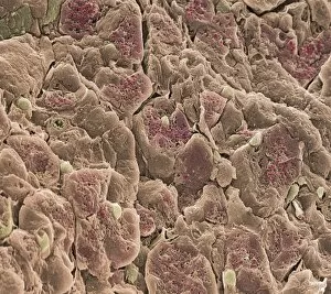
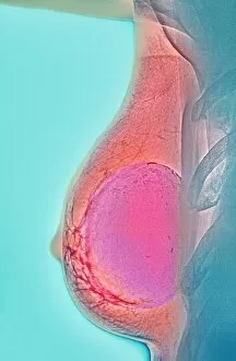
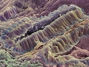
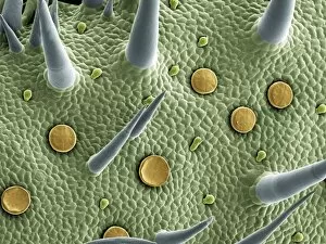
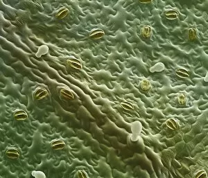
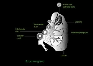
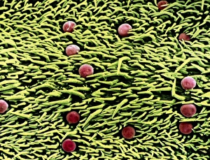
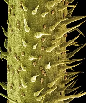
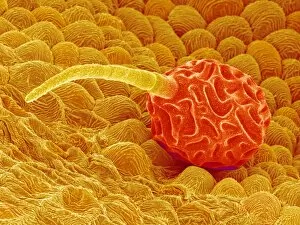
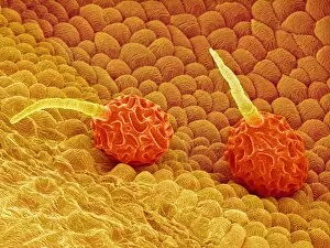
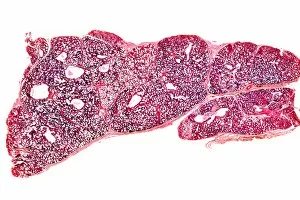
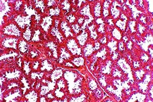
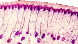
"Glandular: Unveiling the Intricacies of Nature's Secretive World" Botanik Digitalis purpurea L. Fingerhut 160: 1 - A captivating glimpse into the enchanting world of the purple foxglove, revealing its glandular secrets. Exploring Pancreas Anatomy through Artwork - Delving into the intricate structures and functions of this vital organ that plays a crucial role in our body's metabolism. Thyme Leaf Oil Gland - Discovering the hidden treasure within thyme leaves as we unravel the mysteries behind their aromatic essence and medicinal properties. Twinflower (Linnaea borealis) in Flower: Scotland's Rare Gem - Witnessing nature's rarity as we admire the delicate beauty of twinflowers blooming amidst Scotland's breathtaking landscapes. Revealing Glandular Lobelia, Lobelia glandulosa - Uncovering the unique characteristics and therapeutic potential of this fascinating plant species with its distinctive gland-filled blooms. Marveling at Glandular Columbine, Aquilegia glandulosa - Embarking on a journey to explore this stunning flower adorned with exquisite glands that add an extra touch of allure to its appearance. Thyroid Disease: Insights from Baron Jean Louis Alibert's Book - Diving into historical knowledge about thyroid diseases, gaining valuable insights from one of medicine's pioneers who shed light on these conditions centuries ago. Marjoram Leaf Surface under Scanning Electron Microscope (SEM) - Peering through powerful lenses to observe every minute detail on marjoram leaves' surface, unravelling their complex structure and adaptations for survival. Trachea Lining Exposed: SEM C013 / 7126 & C013 / 7122 – Venturing deep inside our respiratory system to examine tracheal linings up close, marveling at their microscopic intricacies and their role in maintaining healthy breathing.