Fraud Blocker Skip to main content

Gastroenteritis Collection

Gastroenteritis, commonly known as the stomach flu

Background imageGastroenteritis Collection: Norovirus particles, TEM

Norovirus particles, TEM
Norovirus particles. Coloured transmission electron micrograph (TEM) of norovirus particles. Norovirus is a genus of RNA (ribonucleic acid)

Background imageGastroenteritis Collection: E. coli bacterium

E. coli bacterium
Escherichia coli. Coloured transmission electron micrograph (TEM) of an Escherichia coli bacterium. E. coli are Gram-negative bacilli (rod-shaped) bacteria

Background imageGastroenteritis Collection: E. coli bacteria

E. coli bacteria
False-colour transmission electron micrograph of the bacterium Escherichia coli, a normal inhabitant of the human intestine

Background imageGastroenteritis Collection: E. coli bacteria, SEM

E. coli bacteria, SEM
E. coli bacteria. Coloured scanning electron micrograph (SEM) of Escherichia coli bacteria (purple) taken from the small intestine of a child. E

Background imageGastroenteritis Collection: E. coli 0157: H7 bacteria

E. coli 0157: H7 bacteria
E. coli 0157:H7 bacteria. Coloured scanning electron micrograph (SEM) of Escherichia coli 0157:H7 bacteria, cause of foodborne illness

Background imageGastroenteritis Collection: Parvovirus particle, artwork C013 / 4640

Parvovirus particle, artwork C013 / 4640
Parvovirus particle. Computer artwork showing the outer structure of a human parvovirus (family Parvoviridae) particle (virion)

Background imageGastroenteritis Collection: Norwalk virus capsid, molecular model F006 / 9372

Norwalk virus capsid, molecular model F006 / 9372
Norwalk virus capsid, molecular model. This norovirus, which causes a viral form of gastroenteritis, is transmitted from person-to-person or through contaminated food

Background imageGastroenteritis Collection: Norwalk virus capsid, molecular model

Norwalk virus capsid, molecular model. This norovirus, which causes a viral form of gastroenteritis, is transmitted from person-to-person or through contaminated food

Background imageGastroenteritis Collection: Parvovirus particles, TEM

Parvovirus particles, TEM
Parvovirus particles. Coloured transmission electron micrograph (TEM) of virus particles (orange) clustered around a piece of cell membrane (purple, centre)

Background imageGastroenteritis Collection: Adenovirus particles and bacterium, TEM

Adenovirus particles and bacterium, TEM
Adenovirus particles and bacterium. Coloured transmission electron micrograph (TEM) of adenovirus particles (green) with a gut bacterium (orange)

Background imageGastroenteritis Collection: Norwalk virus infection

Norwalk virus infection. Computer artwork of a norwalk virus and a human gut. The stomach is at top, the large intestine goes around the outside of the virus

Background imageGastroenteritis Collection: Astrovirus particles, TEM

Astrovirus particles, TEM
Astrovirus particles. Coloured transmission electron micrograph (TEM) of a cluster of astrovirus particles (purple). These are small viruses, measuring approximately 28 nanometres in diameter

Background imageGastroenteritis Collection: Norwalk virus particles

Norwalk virus particles, computer artwork. This small virus consists of an RNA (ribonucleic acid) core surrounded by a protein capsid (multicoloured)

Background imageGastroenteritis Collection: Artwork of digestive system and harmful bacteria

Artwork of digestive system and harmful bacteria
Gastroenteritis. Artwork of bacteria shown below the human digestive system in which they cause gastroenteritis, the inflammation of the stomach and intestines

Background imageGastroenteritis Collection: Norwalk virus particle

Norwalk virus particle. Computer artwork of the capsid of the Norwalk viruswith proteins represented by coloured blobs. The capsid is a protein coat that encloses the viruss RNA (ribonucleic acid)

Background imageGastroenteritis Collection: Coloured TEM of rotaviruses

Coloured TEM of rotaviruses
False-colour transmission electron micrograph (TEM) of rotaviruses. Rotaviruses belong to the genus Reovirus, and are the causative agent of infantile gastroenteritis in humans, mice and calves

Background imageGastroenteritis Collection: Cultured E. coli and Enterococcus bacteria

Cultured E. coli and Enterococcus bacteria
MODEL RELEASED. Hand of a technician holds a petri dish containing agar on which red colonies of Escherichia coli (E.coli)

Background imageGastroenteritis Collection: Petri dish culture of E. coli bacteria

Petri dish culture of E. coli bacteria
MODEL RELEASED. Hand of a technician holds a petri dish containing agar on which black colonies of Escherishia coli (E.coli) bacteria are being cultured in a laboratory. E

Background imageGastroenteritis Collection: Cholera toxin, artwork

Cholera toxin, artwork
Cholera toxin, molecular structure. Cholera is an infectious intestinal disease caused by this toxin produced by the Gram-negative bacterium Vibrio cholerae

Background imageGastroenteritis Collection: Adenovirus particle, TEM

Adenovirus particle, TEM
Adenovirus particle, coloured transmission electron micrograph (TEM). Each particle consists of an icosahedral protein coat (capsid, orange) surrounding double stranded deoxyribonucleic acid (DNA)

Background imageGastroenteritis Collection: E. coli EHEC bacteria, computer artwork

E. coli EHEC bacteria, computer artwork
Computer artwork of a enterohaemorrhagic E. coli (EHEC), a dangerous form of the normally harmless E. coli bacteria which live in the human intestine

Background imageGastroenteritis Collection: Shiga toxin from E. coli

Shiga toxin from E. coli
Computer artwork ribbon diagram of Shiga toxin type 2 (Stx2) from Escherichia coli O157. Shiga toxin is produced by enterohaemorrhagic E. coli (EHEC), a dangerous form of the normally harmless E

Background imageGastroenteritis Collection: Adenovirus particles, TEM

Adenovirus particles, TEM
Adenovirus particles. Coloured transmission electron micrograph (TEM) of adenovirus particles. Each particle consists of an icosahedral protein coat (capsid)

Background imageGastroenteritis Collection: E. coli bacterium strain O157: H7, TEM

E. coli bacterium strain O157: H7, TEM
E. coli bacterium strain O157:H7, coloured transmission electron micrograph (TEM). E. coli are Gram-negative rod-shaped bacteria that are part of the normal flora of the human gut

Background imageGastroenteritis Collection: E. coli bacteria strain O157: H7, TEM

E. coli bacteria strain O157: H7, TEM
E. coli bacteria strain O157:H7, coloured transmission electron micrograph (TEM). E. coli are Gram-negative rod-shaped bacteria that are part of the normal flora of the human gut

Background imageGastroenteritis Collection: Norovirus particles, artwork

Norovirus particles, artwork
Norovirus particles in the gut, artwork. Norovirus is a genus of RNA (ribonucleic acid) viruses that cause about half of all gastroenteritis cases around the world

Background imageGastroenteritis Collection: Neurology research, conceptual artwork

Neurology research, conceptual artwork
Viral research, conceptual computer artwork. Norwalk virus particle (norovirus) being studied under a light microscope. Infection with the Norwalk virus is very common

Background imageGastroenteritis Collection: Norwalk virus contamination, artwork

Norwalk virus contamination, artwork
Norwalk virus contamination, conceptual computer artwork. Door handle contaminated with Norwalk virus, or norovirus, particles (virions)

Background imageGastroenteritis Collection: Salmonella bacterium, TEM

Salmonella bacterium, TEM
Salmonella sp. bacterium, coloured transmission electron micrograph (TEM). Salmonella is a Gram-negative rod-shaped bacterium in the same family as Escherichia coli (Enterobacteriaceae)

Background imageGastroenteritis Collection: Salmonella typhimurium bacterium, SEM

Salmonella typhimurium bacterium, SEM
Salmonella typhimurium bacterium, coloured scanning electron micrograph (SEM). This bacterium is also known as Salmonella enterica serovar typhimurium


All Professionally Made to Order for Quick Shipping

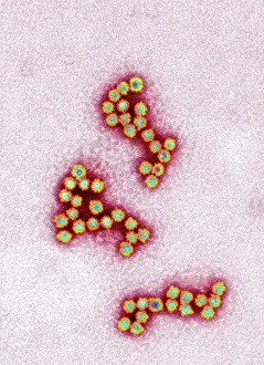
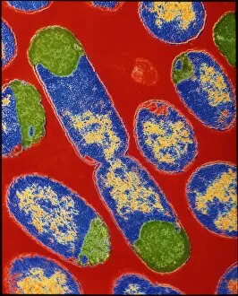
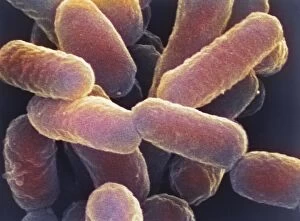
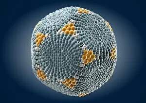
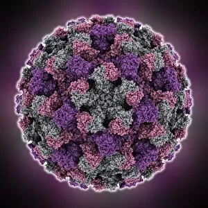
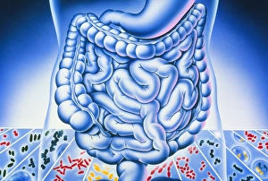
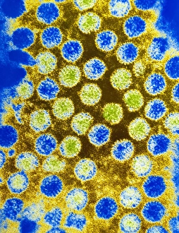
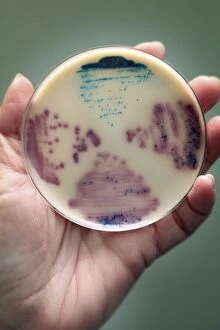
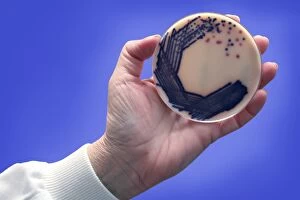
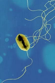
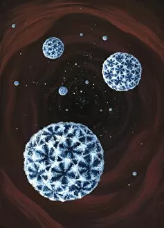
Gastroenteritis, commonly known as the stomach flu, is a highly contagious infection that affects the gastrointestinal tract and can be caused by various pathogens such as Norovirus particles and E. Coli bacteria. When examining Norovirus particles under a transmission electron microscope (TEM), their small size and distinct shape become apparent. These tiny viral particles are responsible for causing outbreaks in crowded places like schools or cruise ships. E. Coli bacterium, specifically E. Coli 0157: H7 bacteria, is another culprit behind gastroenteritis cases. When viewed under a scanning electron microscope (SEM), these bacteria appear rod-shaped with hair-like projections called pili. Some strains of E. coli produce Shiga toxin, which can lead to severe symptoms like bloody diarrhea and kidney damage. Salmonella typhimurium bacteria are also known to cause gastroenteritis when ingested through contaminated food or water. SEM images reveal their unique structure with flagella that help them move around in the digestive system. Escherichia coli bacteria come in different strains and colors, as seen in colored SEM and TEM images. These vibrant visuals highlight the diversity within this bacterial species while emphasizing its role in causing gastrointestinal infections. Understanding the microscopic world of pathogens like Norovirus particles, E. coli bacterium including its harmful strain 0157: H7 along with Salmonella typhimurium bacterium helps us comprehend how these microorganisms contribute to the development - an ailment that affects millions worldwide every year.