
Gamete Collection
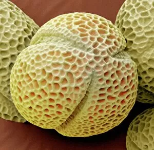
"Unveiling the Intricate World of Gametes: A Closer Look through SEM" Delving into the Microcosm: Exploring Geranium Anther under SEM Captivating Beauty in Detail


All Professionally Made to Order for Quick Shipping
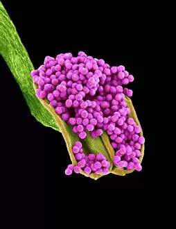
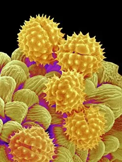
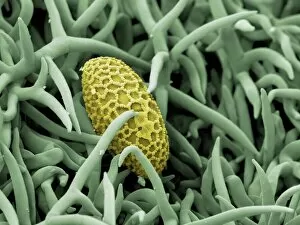
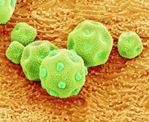
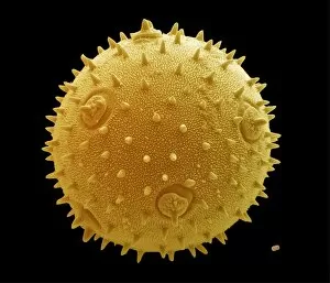
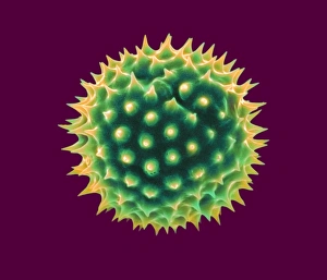
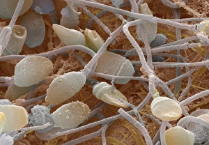
"Unveiling the Intricate World of Gametes: A Closer Look through SEM" Delving into the Microcosm: Exploring Geranium Anther under SEM Captivating Beauty in Detail: Dahlia Flower Pollen Revealed by SEM Passion Unleashed: Witnessing the Exquisite Passion Flower Pollen via SEM Honeybee's Secret Treasure: Discovering Pollen on its Leg with SEM The Marvels of Reproduction: Geranium Pollen Magnified under SEM Life Begins to Flourish: Embryo Development 24-36 Hours after Fertilization, a Mesmerizing Journey Contraception and Sperm Cells Collide: IUD as a Barrier against Fertilization Explored Nature's Tiny Warriors Unveiled: Dandelion Pollen Grain Examined through SEM Hitchhiking on Bees' Journeys: Tracing Pollen Grains on Bee Legs with SEM Philadelphia Fleabane's Hidden Gems Revealed by SEM - Stunningly Beautiful Pollen Grains Up Close Gorse Stigma Embraces Life's Essence - Glimpsing at Its Adorned Surface with Polished Pollens Underneath, Captured by anSEM Lens A Serendipitous Encounter - Lily pollen grain finds solace upon Rosemary leaf.