
Frontal Lobe Collection
"The Frontal Lobe: Unveiling the Command Center of the Human Brain" The frontal lobe, a vital component of the intricate anatomy of the human brain


All Professionally Made to Order for Quick Shipping
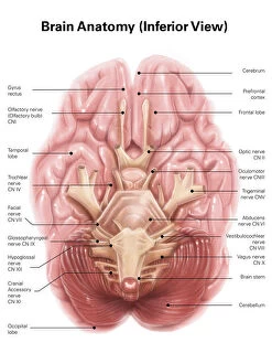
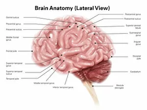
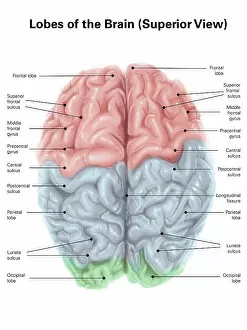
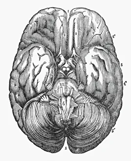
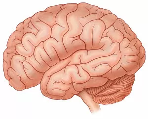
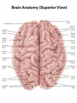
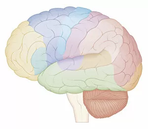
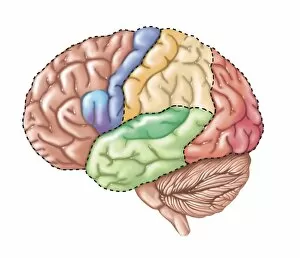
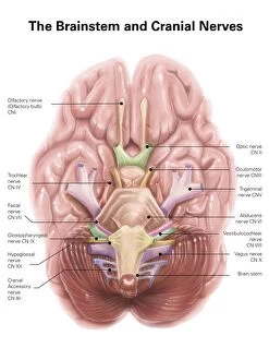
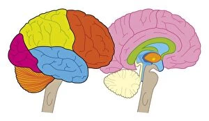
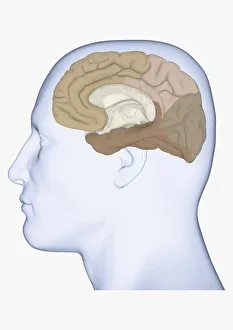
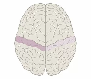
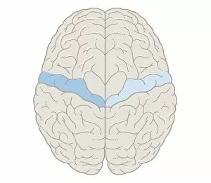
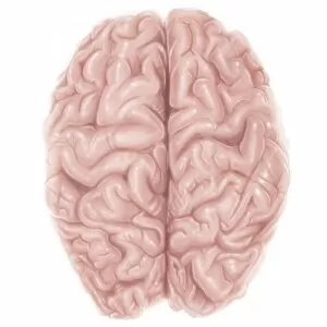
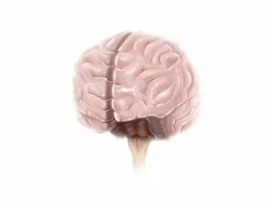
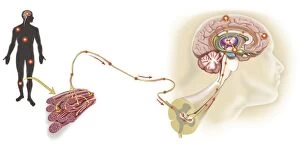
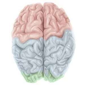
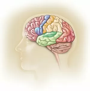
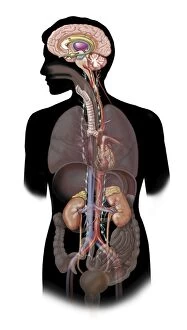
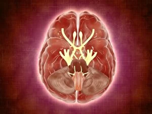
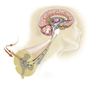
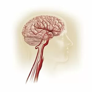
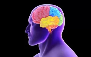
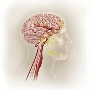
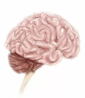
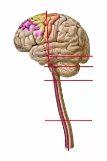
"The Frontal Lobe: Unveiling the Command Center of the Human Brain" The frontal lobe, a vital component of the intricate anatomy of the human brain, takes center stage in our cognitive abilities and decision-making processes. From its commanding position at the front and top region of our brain, this remarkable structure plays a crucial role in shaping who we are. Inferior view: As we explore beneath the surface, an inferior view reveals how this cerebral powerhouse connects to other lobes and structures within our brain. It serves as a bridge between different regions, allowing for seamless communication and coordination. Superior view with colored lobes: A superior view showcases this magnificent organ adorned with vibrant colors that highlight its distinct lobes. The frontal lobe stands out prominently among them all, symbolizing its prominence in governing various functions such as reasoning, problem-solving, planning, and personality expression. Lateral view: Observing from a lateral perspective unveils another dimension of complexity within the frontal lobe's architecture. This angle allows us to appreciate its interconnectedness with neighboring areas like basal ganglia – an artistic representation showcasing their interplay in motor control and reward mechanisms. Under Surface Engraved Illustration (Circa 1880): Delving into history through an engraved illustration from 1880 offers a glimpse into how early anatomists meticulously captured the under-surface details of the human brain. Such depictions remind us of humanity's enduring fascination with unraveling these mysteries throughout time. Normal coronal section: A normal coronal section provides insight into both skull and brain anatomy while highlighting coronal sinuses' presence. Within this cross-section lies one of nature's most extraordinary creations – where intelligence meets resilience. Superior View Biomedical Illustration: In a superior view biomedical illustration, we witness intricate neural pathways intricately mapped across every inch of this awe-inspiring organ. These pathways form networks responsible for transmitting information, orchestrating our thoughts, emotions, and actions.