
Flagellate Collection
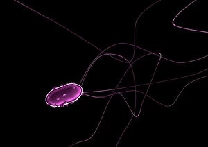
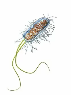
"Exploring the World of Flagellate: From Bacteria to Artwork and Beyond" Flagellate bacteria


All Professionally Made to Order for Quick Shipping
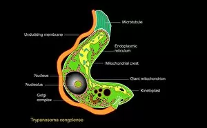
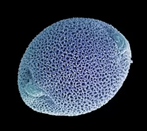
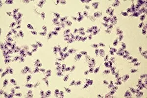
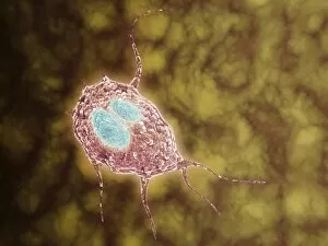
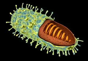
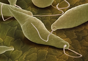
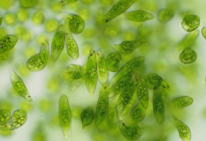
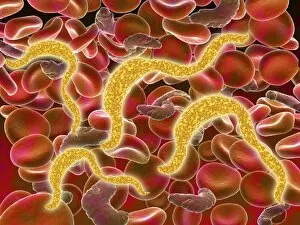
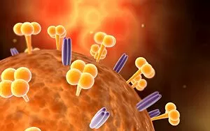
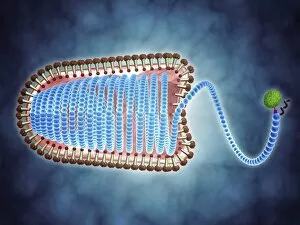
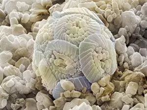
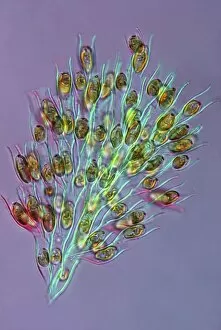
"Exploring the World of Flagellate: From Bacteria to Artwork and Beyond" Flagellate bacteria: These tiny organisms propel themselves using whip-like structures called flagella, enabling them to move through various environments. Calcareous phytoplankton fossil, SEM Z100 / 0213: This scanning electron microscope image reveals the intricate details of a fossilized calcareous phytoplankton, showcasing their unique flagellated structures. Trypanosome protozoan, artwork: Through artistic interpretation, this artwork captures the mesmerizing beauty of a trypanosome protozoan with its distinct flagella that aid in locomotion. Dinoflagellates, SEM: In this stunning scanning electron microscope image, dinothey are showcased with their characteristic flagella that contribute to their diverse ecological roles in marine ecosystems. TEM of Trichomonas vaginalis: Transmission electron microscopy provides an up-close view of Trichomonas vaginalis, a parasitic protozoan known for its multiple flagella that enable it to move within the human reproductive system. False-color TEM of Salmonella typhi: This false-color transmission electron micrograph highlights the presence of multiple flagella on Salmonella typhi bacteria responsible for causing typhoid fever. The Flagellation (1512): A burin engraving on copper from 1512 depicts the biblical scene known as "The Flagellation, " illustrating Christ's suffering and punishment before his crucifixion. The Flagellation (c. 1395-1400): Painted with tempera on canvas and adorned with gold ground accents, this masterpiece showcases "The Flagellation" scene from medieval times—depicting Christ's torment during his Passion. The Flagellation of Christ (polychrome wood).