
Filiform Collection
"Exploring the Intricate World Papillae: Unveiling the Secrets of Taste Sensation" In this captivating journey


All Professionally Made to Order for Quick Shipping
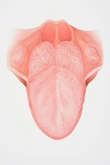
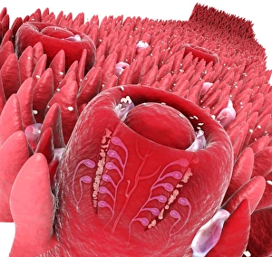
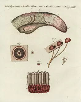
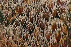
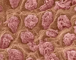
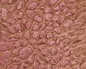
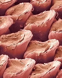
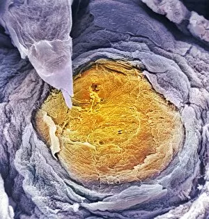
"Exploring the Intricate World Papillae: Unveiling the Secrets of Taste Sensation" In this captivating journey, we delve into the fascinating world papillae – tiny structures that play a crucial role in our sense of taste. Through various microscopic views and illustrations, we unravel the intricate anatomy and function of these remarkable features found on our tongues. A front view diagram illustrates the complexity of tongue anatomy, highlighting the presence papillae among other types. These slender projections are further showcased through digital cross-section illustrations, revealing their close association with taste buds. Zooming in even closer, we observe a cow tongue under a microscope, where an array papillae covers its surface like delicate bristles. Similarly, human tongues come into focus through scanning electron microscopy (SEM), showcasing their unique texture and arrangement papillae. Not limited to humans alone, cat tongues also exhibit intriguing patterns when observed at high magnification using SEM. The detailed images highlight how these tiny structures differ across species while serving similar purposes. Delving deeper into micrographs taken under light microscopy reveals stunning details about tongue papillae's cellular composition. These images provide insights into their structural intricacies and underline their vital role in enhancing our gustatory experiences. Lastly, SEM captures tantalizing glimpses inside taste buds themselves – specialized sensory organs responsible for detecting different flavors. By examining these microscopic marvels alongside surrounding filiform papillae, scientists continue to uncover secrets behind our ability to savor diverse tastes. Through this visual exploration encompassing multiple perspectives and techniques, we gain newfound appreciation for the often-overlooked yet indispensable role played by filiform papillae in shaping our perception of flavor.