
Fibre Collection (page 7)
"Fibre: Unveiling the Intricacies of Connectivity and Strength" Delving into the depths of our complex neural network, brain fibres emerge as the architects of cognition


All Professionally Made to Order for Quick Shipping
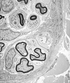
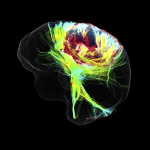
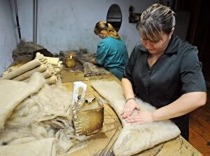
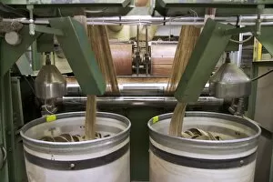
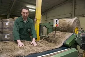
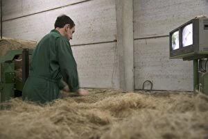
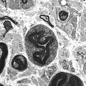
"Fibre: Unveiling the Intricacies of Connectivity and Strength" Delving into the depths of our complex neural network, brain fibres emerge as the architects of cognition. Revealed through DTI MRI scans such as C017/7099 and C017/7035, these intricate pathways illuminate the wonders within. Just like a Ducati 998R from Italy roars with power on the racetrack, brain fibres propel our thoughts at lightning speed. They navigate through convoluted terrains, connecting different regions to orchestrate seamless communication. In an unexpected twist, mixing treated asbestos fibre becomes a spectacle when paired with Heath Robinson's ingenious machine. This patent double action grinder showcases human ingenuity in harnessing strength even from unlikely sources. While Mercedes-Benz SL65 AMG Black Series exudes elegance on wheels, it is woven fabric under SEM that captivates us with its delicate intricacy. Each thread weaves together to create a tapestry of resilience and beauty. Juicing carrots reveals another facet of fibre's significance – nourishment for our bodies. As citrus fruits burst with tangy flavors, their fibrous content fortifies our health while adding zest to life itself. Examining a lime tree stem under light micrograph unravels nature's own masterpiece – an intricate network designed for growth and survival. It reminds us that even in simplicity lies extraordinary complexity waiting to be discovered. Myelination of nerve fibres captured by TEM showcases how protective layers enhance conductivity within our nervous system. Like armor shielding warriors in battle, myelin ensures swift transmission and efficient functioning. From brain fibers orchestrating thoughts to treating asbestos fiber ingeniously or marveling at woven fabrics' artistry - each instance highlights fiber’s versatility across various domains. Whether powering machines or nurturing bodies, fiber intertwines seamlessly into every aspect of life - connecting us all in ways unseen yet indispensable.