Fraud Blocker Skip to main content

Feeding Structure Collection

"Exploring the Intricate Feeding Structures of Insects

Background imageFeeding Structure Collection: Female mosquito, SEM

Female mosquito, SEM
Female mosquito. Coloured scanning electron micrograph (SEM) of the underside of an unidentified female mosquito (family Culicidae). The large compound eyes (pink) dominate the head

Background imageFeeding Structure Collection: Mosquito larva head

Mosquito larva head. Coloured scanning electron micrograph (SEM) of the head of an unidentified mosquito larva (family Culicidae)

Background imageFeeding Structure Collection: Flys proboscis, SEM

Flys proboscis, SEM
Flys proboscis. Coloured scanning electron micrograph (SEM) of the tip of the proboscis (snout) of an unidentified fly (order Diptera). Flies possess mouthparts adapted for piercing and sucking

Background imageFeeding Structure Collection: Mosquito larva mouth hairs

Mosquito larva mouth hairs. Coloured scanning electron micrograph (SEM) of the mouth hairs of a mosquito larva (Aedes albopictus). These hairs are feeding structures used to filter water

Background imageFeeding Structure Collection: Flys proboscis, light micrograph

Flys proboscis, light micrograph
Flys proboscis. Light micrograph (LM) of the tip of the proboscis (snout) of a fly (order Diptera). Flies possess mouthparts adapted for piercing and sucking

Background imageFeeding Structure Collection: Hover fly proboscis, SEM

Hover fly proboscis, SEM
Hover fly proboscis, coloured scanning electron micrograph (SEM). This flys mouthparts are adapted for piercing and sucking

Background imageFeeding Structure Collection: Fly proboscis, SEM

Fly proboscis, SEM
Fly proboscis. Coloured scanning electron micrograph (SEM) of the tip of the proboscis (snout) of an unidentified fly (order Diptera). Flies possess mouthparts adapted for piercing and sucking

Background imageFeeding Structure Collection: Hydrozoan colony

Hydrozoan colony. Close-up of the tentacled feeding structures of a hydrozoan (Tubularia larynx) colony. Hydrozoa are a type of cnidarian and are related to corals and sea anemones

Background imageFeeding Structure Collection: Hydrozoan colony and amphipods

Hydrozoan colony and amphipods. Amphipod (Stenothoe marina) crustaceans sheltering amongst the tentacled feeding structures of a hydrozoan (Tubularia larynx) colony

Background imageFeeding Structure Collection: Hydrozoan

Hydrozoan (Tubularia indivisa). Hydrozoa are a type of cnidarian and are related to corals and sea anemones


All Professionally Made to Order for Quick Shipping

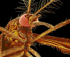
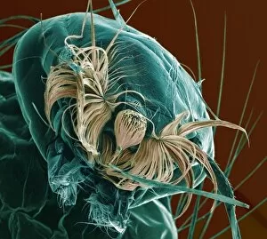
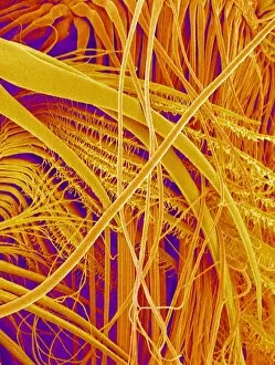
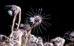
"Exploring the Intricate Feeding Structures of Insects: A Close-up Look through SEM and Light Micrograph" Insects possess fascinating feeding structures that allow them to extract nutrients in unique ways. Through the use of advanced imaging techniques such as Scanning Electron Microscopy (SEM) and light micrographs, we can delve into the intricate details of these feeding mechanisms. One such example is the fly proboscis, a long tubular mouthpart used for sipping nectar or other liquids. Under SEM, this delicate structure reveals its fine grooves and tiny hairs that aid in fluid uptake. Similarly, female mosquitoes exhibit a specialized proboscis designed for piercing skin and drawing blood. SEM images showcase its sharp needle-like structure with serrated edges, enabling efficient blood-feeding. Mosquito larvae also display intriguing feeding adaptations. The head region of these larvae exhibits distinct features under SEM, including sensory organs and mouthparts adapted for filter-feeding on organic matter present in water bodies. The mosquito larva's mouth hairs play a crucial role in capturing food particles from their aquatic environment. These microscopic bristles are visible under high-resolution microscopy images, highlighting their importance in nutrient acquisition during early life stages. Not limited to flies and mosquitoes alone, hoverflies also possess remarkable proboscises tailored for specific feeding habits. SEM imagery allows us to observe the complex arrangement of bristles on hoverfly proboscises which aid them in accessing floral nectar efficiently. By studying these diverse insect species' feeding structures using cutting-edge imaging technologies like SEM and light micrographs, scientists gain valuable insights into their ecological roles and evolutionary adaptations. Understanding these intricate mechanisms not only deepens our knowledge but also provides inspiration for biomimetic designs aimed at improving various aspects of human life.