Fraud Blocker Skip to main content

False Colour Collection (page 16)

"Unveiling the Hidden Beauty: Exploring False Colour in Microscopy and Imaging" Step into a world where reality meets artistry

Background imageFalse Colour Collection: Salivary gland, light micrograph

Salivary gland, light micrograph
Salivary gland. Coloured light micrograph of a section through a sublingual salivary gland, which is situated directly under the tongue

Background imageFalse Colour Collection: Tongue papillae, light micrograph

Tongue papillae, light micrograph
Tongue papillae. Coloured light micrograph of a section through the tongue, showing its filiform papillae (red). They project from the tongues upper surface, giving it its rough texture

Background imageFalse Colour Collection: Taste bud, light micrograph

Taste bud, light micrograph
Taste bud. Coloured light micrograph of a section through the tongue, showing a taste bud. This taste bud is seen as a round purple mass slightly to the right of the vertical white line

Background imageFalse Colour Collection: Eye structure, light micrograph

Eye structure, light micrograph
Eye structure. Coloured light micrograph of a section through the inner surface of a human eye. This slice shows the vitreous humour (transparent liquid in the eye, top), the retina (pink)

Background imageFalse Colour Collection: Vision and the brain, MRI scan

Vision and the brain, MRI scan
Vision and the brain. Coloured Magnetic Resonance Imaging (MRI) scan of an axial section through the head of a 55 year old man, showing the eyes, optic nerves and brain

Background imageFalse Colour Collection: The iris of the eye

The iris of the eye
Iris. Computer-enhanced image of the blue/brown iris of a human eye. The iris is a ring of muscle fibres which contracts and relaxes to alter the light entering the eye through the pupil (black)

Background imageFalse Colour Collection: Iris

Iris. Computer-enhanced and coloured image of a human iris. The iris is a ring of muscle fibres. It contracts and relaxes to alter the amount of light entering the eye through the pupil (black)

Background imageFalse Colour Collection: Myelinated nerve fibres, SEM

Myelinated nerve fibres, SEM
Myelinated nerve fibres. Coloured scanning electron micrograph (SEM) of a section through a bundle of nerve fibres containing a blood vessel (orange)

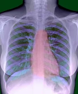
Background imageFalse Colour Collection: Heart, chest X-ray

Heart, chest X-ray

Background imageFalse Colour Collection: Lymph node, light micrograph

Lymph node, light micrograph
Lymph node. Coloured light micrograph of a section through a lymph node. A lymph node filters pathogens from lymph fluid, a watery liquid that surrounds the tissues of the body

Background imageFalse Colour Collection: Macrophage cell, TEM

Macrophage cell, TEM
Macrophage. Coloured transmission electron micrograph (TEM) of a macrophage cell. The cells nucleus is bright green. Mitochondria (green) in the cells cytoplasm produce energy for the cell

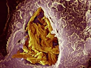
Background imageFalse Colour Collection: Macrophage engulfing bacteria, SEM

Macrophage engulfing bacteria, SEM
Macrophage engulfing bacteria. Coloured scanning electron micrograph (SEM) of a macrophage white blood cell (brown) engulfing Mycobacterium tuberculosis bacteria (yellow)

Background imageFalse Colour Collection: Macrophage engulfing tuberculosis vaccine

Macrophage engulfing tuberculosis vaccine. Coloured scanning electron micrograph (SEM) of a macrophage white blood cell engulfing Mycobacterium bovis bacteria (yellow)

Background imageFalse Colour Collection: Neutrophil cell and bacteria, SEM

Neutrophil cell and bacteria, SEM
Neutrophil cell and bacteria. Coloured scanning electron micrograph (SEM) of bacteria (rod-shaped) and part of a neutrophil cell (lower left)

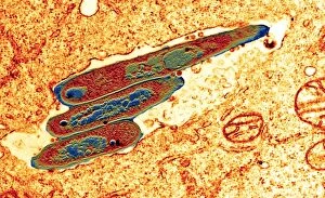
Background imageFalse Colour Collection: Macrophage cell engulfing bacteria, TEM

Macrophage cell engulfing bacteria, TEM
Macrophage cell engulfing bacteria. Coloured transmission electron micrograph (TEM) of bacteria (centre, rod-shaped) inside a macrophage cell

Background imageFalse Colour Collection: Blood clot crystals, SEM

Blood clot crystals, SEM
Blood clot crystals. Coloured scanning electron micrograph (SEM) of crystals of albumin from a blood clot. Albumin is the most abundant protein in the blood plasma

Background imageFalse Colour Collection: Human antibodies, TEM

Human antibodies, TEM
Human antibodies (yellow), coloured transmission electron micrograph (TEM). The Y-shaped structures are molecules of the immunoglobulin G (IgG) antibody

Background imageFalse Colour Collection: Red blood cells, SEM

Red blood cells, SEM
Red blood cells. Coloured scanning electron micrograph (SEM) of red blood cells (erythrocytes). Red blood cells are biconcave, disc-shaped cells that transport oxygen from the lungs to body cells


All Professionally Made to Order for Quick Shipping

"Unveiling the Hidden Beauty: Exploring False Colour in Microscopy and Imaging" Step into a world where reality meets artistry, as we delve into the captivating realm of "false colour. " In this mesmerizing journey, we encounter an array of intricate subjects that have been transformed through advanced imaging techniques. Brace yourself for a visual feast that transcends our conventional perception. First up, behold the brain's intricate network of blood vessels captured in a 3D angiogram from 1981. Through false colour enhancement, these delicate pathways come alive with vibrant hues, revealing their hidden complexity. Moving on to nature's wonders, prepare to be enchanted by the Geranium anther under scanning electron microscopy (SEM). Its microscopic details are unveiled in vivid shades, showcasing its remarkable structure and beauty. Next on our exploration is Dahlia flower pollen also observed under SEM. The once minuscule grains now emerge as magnificent orbs adorned with striking colours—a testament to nature's exquisite design. Venturing further into the microscopic realm, we encounter diatoms—tiny algae—with their intricately patterned shells. Under SEM and enhanced with false colouring techniques, these diatoms transform into kaleidoscopic masterpieces that blur the line between science and art. But it doesn't stop there. Witness an ant like never before through SEM; its exoskeleton becomes a work of art when illuminated by vibrant tones. Meanwhile, X-ray imaging reveals the intricate skeleton of a triggerfish—an ethereal display reminiscent of otherworldly creatures lurking beneath ocean depths. Delving deeper within cells brings us face-to-face with rough endoplasmic reticulum—the cellular factory responsible for protein synthesis. Enhanced by false colouring using transmission electron microscopy (TEM), this complex network takes on an otherworldly appearance akin to futuristic cityscapes bathed in neon lights. Shifting gears towards medical marvels captured via X-ray technology unveils both the functional and aesthetic aspects of our bodies.