
Er Collection (page 3)
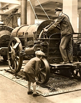
"Exploring the 'er' in History, Science, and Art" George Stephenson (1781-1848): A pioneer in locomotive engineering


All Professionally Made to Order for Quick Shipping
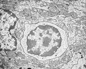
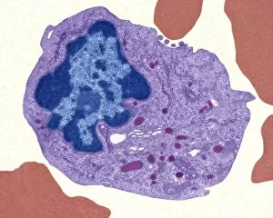
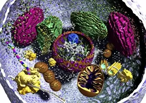
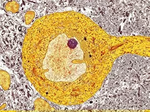
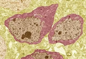
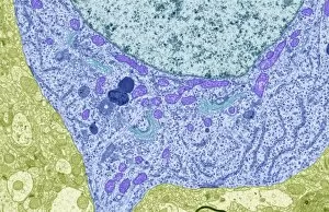
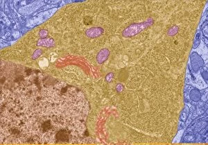
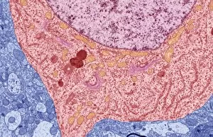
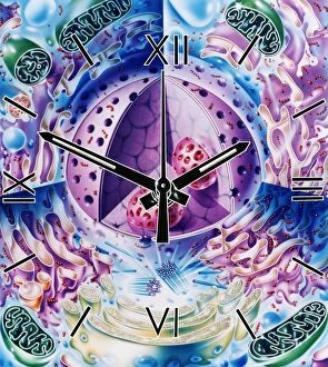
"Exploring the 'er' in History, Science, and Art" George Stephenson (1781-1848): A pioneer in locomotive engineering, George Stephenson revolutionized transportation with his innovative designs. Rough endoplasmic reticulum, TEM: This microscopic image reveals the intricate network of membranes within cells responsible for protein synthesis and transport. Trypanosome protozoan, artwork: Delve into the world of microorganisms through captivating artwork depicting a trypanosome protozoan—a parasite causing diseases like sleeping sickness. George Stephenson's Hetton colliery locomotive: Witness the power of steam as you admire this historic locomotive that once transported coal from mines to industrial centers. Aberystwith, c1900. Artist: ER Gyde: Immerse yourself in a picturesque scene captured by artist ER Gyde, showcasing the beauty of Aberystwith during the early 20th century. Purkinje nerve cell, TEM C014 / 0583: Peer into the complexity of our nervous system with an electron microscope image revealing Purkinje cells—key players in coordinating movement and balance. Fibroblast cell, artwork: Marvel at an artistic representation highlighting fibroblast cells—the building blocks of connective tissue essential for wound healing and maintaining tissue integrity. Locomotive passenger engine no 73: Step back in time as you gaze upon this vintage locomotive that once transported passengers across vast landscapes with elegance and efficiency. A postbox in snow UK: Capture a serene winter moment through this photograph featuring a red postbox standing tall amidst a snowy landscape—an iconic symbol of communication even during harsh weather conditions. Maritime - brass gimble lamp: Illuminate your imagination with a maritime-themed brass gimble lamp—a timeless piece used aboard ships to provide steady light despite rough seas or turbulent voyages.