
Epithelial Cells Collection
Epithelial cells, the building blocks of our body's tissues and organs, come in various shapes and sizes


All Professionally Made to Order for Quick Shipping
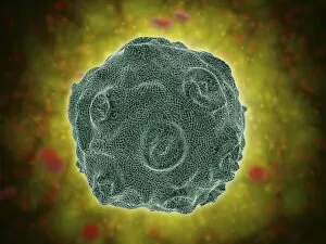
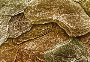
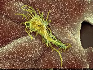
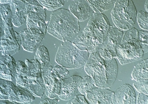
Epithelial cells, the building blocks of our body's tissues and organs, come in various shapes and sizes. In a cytologic smear of the vagina, we can observe intermediate squamous cells that line this important reproductive organ. These cells play a crucial role in maintaining its health and functionality. One significant aspect related to epithelial cells is their susceptibility to infections caused by Human Papilloma Virus (HPV). A conceptual image depicting this virus serves as a reminder of the potential risks associated with unprotected sexual activity. Moving on to different types of epithelia, simple cuboidal epithelium is represented by another conceptual image. This type of tissue forms small ducts within glands or kidney tubules, aiding in secretion and absorption processes. Stratified squamous epithelium is yet another variant showcased through an illustrative representation. Found in areas subjected to wear and tear like the skin or esophagus lining, it acts as a protective barrier against external factors such as pathogens or physical damage. In contrast, simple columnar epithelium provides specialized functions within our digestive system. The conceptual image highlights its presence along the inner lining of organs like the stomach or intestines where it aids in nutrient absorption. Pseudostratified columnar epithelium presents itself as an intriguing variation captured through visual imagery. Despite appearing stratified due to nuclei at varying heights, all these cells actually touch the basement membrane beneath them. This tissue lines parts of our respiratory tract and helps trap foreign particles from entering deeper into our lungs. Delving further into anatomical structures, we encounter an illustration showcasing the intricate layers comprising the stomach wall. Epithelial cells form one layer among others responsible for digestion-related activities occurring within this vital organ. The diversity continues with stratified cuboidal epithelium depicted conceptually; this type typically lines larger ducts found in sweat glands or mammary glands during lactation periods.