Fraud Blocker Skip to main content

Epithelia Collection

Epithelia, the microscopic heroes of our body's defense system

Background imageEpithelia Collection: Gallbladder surface, SEM C015 / 9613

Gallbladder surface, SEM C015 / 9613
Gallbladder surface, coloured scanning electron micrograph (SEM). The columnar epithelium of the surface is covered in microvilli (tiny hair-like projections)

Background imageEpithelia Collection: Gallbladder surface, SEM C015 / 9611

Gallbladder surface, SEM C015 / 9611
Gallbladder surface, coloured scanning electron micrograph (SEM). At centre are three secretory glands (yellow). The columnar epithelium of the surface is covered in microvilli

Background imageEpithelia Collection: Gallbladder surface, SEM C015 / 9612

Gallbladder surface, SEM C015 / 9612
Gallbladder surface, coloured scanning electron micrograph (SEM). The columnar epithelium of the surface is covered in microvilli (tiny hair-like projections)

Background imageEpithelia Collection: Kidney tubule, TEM

Kidney tubule, TEM
Kidney tubule. Coloured transmission electron micrograph (TEM) of a section through a proximal convoluted tubule in the kidney

Background imageEpithelia Collection: Kidney tubules, SEM

Kidney tubules, SEM
Kidney tubules. Coloured scanning electron micrograph (SEM) of proximal convoluted tubules (dark brown) in the kidney. The proximal convoluted tubules function to reabsorb water

Background imageEpithelia Collection: Nasal epithelial cells, TEM

Nasal epithelial cells, TEM
Nasal epithelial cells. Coloured transmission electron micrograph (TEM) of stratified squamous epithelial cells from the lining of the nasal cavity

Background imageEpithelia Collection: Blood vessel and cells, computer artwork

Blood vessel and cells, computer artwork
Blood vessel and cells. Computer artwork of various blood cells and a blood vessel. The blood vessel consists of an outer layer of muscle (pink)

Background imageEpithelia Collection: Kidney tubules

Kidney tubules. Coloured scanning electron micrograph (SEM) of proximal convoluted tubules (dark brown) in the kidney. The proximal convoluted tubules function to reabsorb water

Background imageEpithelia Collection: Human epithelial cells

Human epithelial cells. Fluorescent light micrograph of human epithelial cells in culture. The nuclei, which contain the cells genetic information DNA (deoxyribonucleic acid), have been dyed blue

Background imageEpithelia Collection: Eye lens cells, SEM

Eye lens cells, SEM
Eye lens. Coloured scanning electron micrograph (SEM) of a freeze-fractured lens (crystalline lens) of an eye, showing stacked cuboidal epithelium cells

Background imageEpithelia Collection: Fallopian tube, TEM

Fallopian tube, TEM
Fallopian tube, coloured transmission electron micrograph (TEM). Section through non-ciliated columnar epithelium from a fallopian tube. The ciliated cells are not shown here

Background imageEpithelia Collection: Trachea mucous membrane, SEM

Trachea mucous membrane, SEM
Trachea mucous membrane. Coloured scanning electron micrograph (SEM) of a fractured mucous membrane of the trachea (wind pipe), showing the epithelium and underlying connective tissue

Background imageEpithelia Collection: Fallopian tube cells, SEM

Fallopian tube cells, SEM
Fallopian tube cells. Coloured scanning electron micrograph (SEM) of the surface of a fallopian tube, showing non-ciliated (pink) and ciliated (yellow) cells

Background imageEpithelia Collection: Bile duct, SEM

Bile duct, SEM
Bile duct. Coloured scanning electron micrograph (SEM) of a fractured bile duct. Columnar epithelial cells are red and brown, lamina propria is green and microvilli are purple

Background imageEpithelia Collection: Skin, SEM

Skin, SEM
Skin. Coloured scanning electron micrograph (SEM) of squamous epithelial cells on the skin surface. Magnification: x350 when printed at 10 centimetres wide

Background imageEpithelia Collection: Gall bladder surface, SEM

Gall bladder surface, SEM
Internal lining of the gall bladder, coloured scanning electron micrograph (SEM). These columnar epithelial cells are covered in microvilli. Magnification: x2100 when printed at 10 centimetres wide

Background imageEpithelia Collection: Intestinal papillomas, SEM

Intestinal papillomas, SEM
Intestinal papillomas. Coloured scanning electron micrograph (SEM) of a papilloma from a persons intestine. Papillomas are benign (non-cancerous) tumours that arise from epithelial (lining) tissue

Background imageEpithelia Collection: Fallopian tube, SEM

Fallopian tube, SEM
Fallopian tube. Coloured scanning electron micrograph (SEM) of the lining of a fallopian tube (oviduct). The fallopian tubes carry the egg from the ovary to the uterus (womb)

Background imageEpithelia Collection: Nasal epithelium, SEM

Nasal epithelium, SEM
Nasal epithelium, coloured scanning electron micrograph (SEM). The nasal cavity contains both cilated (pink) and non-ciliated (secretory) cells

Background imageEpithelia Collection: Bacteria in the nose, SEM

Bacteria in the nose, SEM
Bacteria in the nose. Coloured scanning electron micrograph (SEM) of bacteria (red) on the surface of the nasal cavity. One of the squamous epithelium cells (lower centre to lower right)

Background imageEpithelia Collection: Oviduct mucosal folds, light micrograph

Oviduct mucosal folds, light micrograph. This section through the folds shows the ciliated columnar epithelium (red) and the connective tissue core (green)


All Professionally Made to Order for Quick Shipping

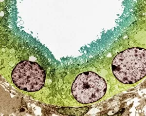
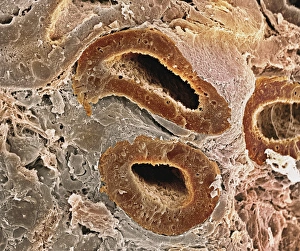
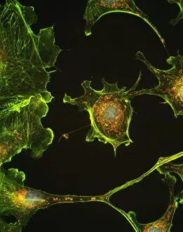
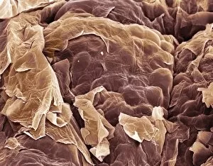
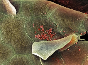
Epithelia, the microscopic heroes of our body's defense system. 🌟 From the bacteria-fighting warriors in our nose to the intricate structures within our organs, epithelial cells play a vital role in maintaining our health. Let's take a closer look at these fascinating entities through various scanning and transmission electron microscopy (SEM and TEM) images. In one captivating SEM image, we observe bacteria residing on the surface of nasal epithelial cells. These tiny organisms are met with fierce resistance from these specialized cells, which act as guardians against invading pathogens. Shifting focus to another SEM capture, we explore the gallbladder surface. The highly detailed C015/9613 image showcases its unique architecture, revealing an intricate network of epithelial cells that line this organ. Their arrangement ensures efficient bile storage and release for digestion purposes. Continuing our exploration of gallbladder surfaces through SEM imagery, we encounter two more stunning captures labeled C015/9611 and C015/9612. These visuals provide further insight into the delicate structure of epithelial cells found here – their interconnections forming a protective barrier while facilitating essential functions. Moving deeper into our internal systems using TEM imaging techniques, we delve into kidney tubules' microscopic world. Two TEM images showcase these intricate structures responsible for filtering waste products from blood while reabsorbing valuable substances like water and electrolytes. TEM also allows us to zoom in on cell nuclei – command centers orchestrating cellular activities. A striking TEM visualization presents us with this central hub brimming with genetic information inside a kidney tubule cell nucleus. Stepping away from kidneys but staying within reproductive anatomy territory, an intriguing SEM image reveals fallopian tube epithelium. This delicate tissue plays a crucial role in guiding eggs towards fertilization sites within the female reproductive system. Returning to TEM imaging once again brings us face-to-face with another glimpse into cell nuclei; this time observed within nasal epithelial cells.