
Enteric Collection
"Exploring the Intricate World of Enteric: Unveiling the Microscopic Marvels and Historical Connections" Delving into the microscopic realm, we encounter E


All Professionally Made to Order for Quick Shipping
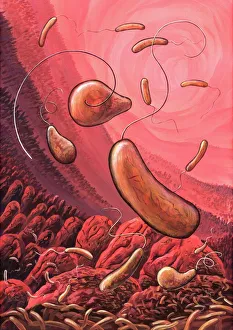
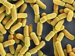
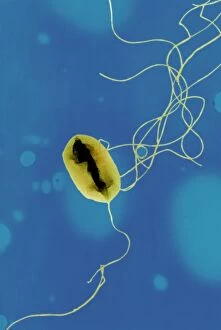
"Exploring the Intricate World of Enteric: Unveiling the Microscopic Marvels and Historical Connections" Delving into the microscopic realm, we encounter E. Coli bacteria through scanning electron microscopy (SEM), revealing their intricate structures. These tiny organisms, often associated with gastrointestinal infections, captivate scientists with their complex morphology. Transitioning to transmission electron microscopy (TEM), we observe a single E. Coli bacterium in astonishing detail. Its delicate features come to life under high magnification, showcasing its unique characteristics and providing valuable insights for researchers. Shifting gears from microorganisms to historical figures, we stumble upon an intriguing connection between Princess Helena and Prince Christian Victor alongside our exploration wonders. Their presence adds a touch of royalty to this captivating journey. Artwork depicting cholera bacteria catches our attention next—a reminder of the devastating impact that enteric diseases have had throughout history. The artwork serves as a poignant representation of humanity's ongoing battle against these formidable foes. Returning to SEM imagery, we once again encounter E. Coli bacteria—this time displaying different strains denoted by C014 / 0385 and C014 / 0386 codes. Each strain possesses its own distinct characteristics, highlighting the vast diversity within this microbial world. Lactobacillus bacteria make their appearance under SEM as well—an important counterbalance in maintaining gut health amidst potential pathogenic threats like E. coli. These beneficial microbes play a crucial role in digestion and overall well-being. Amidst our scientific expedition lies an unexpected historical gem—the paragon prince Henry, eldest son of James I & VII—whose legacy intertwines with our exploration wonders across time. TEM reveals another fascinating aspect—the myenteric nerve plexus intricately woven within the gut wall's smooth muscle fibers—a vital component ensuring proper digestive function and communication between neurons along the intestinal tract. Concluding our captivating journey through enteric marvels, we once again encounter the E.