
Endocrine Collection
The endocrine system, a complex network of glands and hormones, plays a vital role in maintaining homeostasis within the human body


All Professionally Made to Order for Quick Shipping
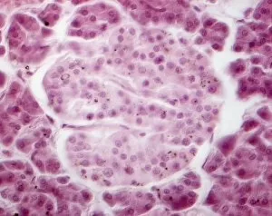
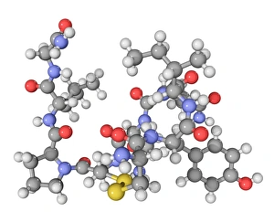
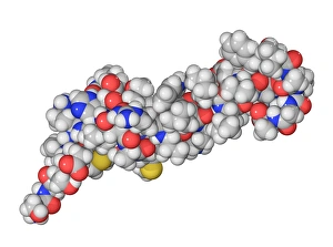
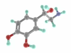
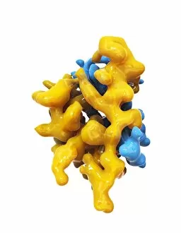
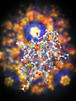
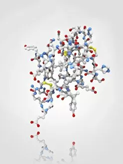
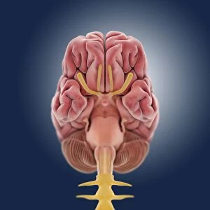
The endocrine system, a complex network of glands and hormones, plays a vital role in maintaining homeostasis within the human body. From ancient times to modern science, the study disorders has fascinated researchers and artists alike. In the 15th century artwork C018/1849, goitre is depicted as an enlargement of the thyroid gland due to iodine deficiency. This condition highlights the importance of proper nutrition for healthy hormone production. Pancreas anatomy is beautifully illustrated in another artwork, showcasing its intricate structure. The pancreas houses the Islet of Langerhans, which can be observed under a light micrograph. These tiny clusters produce insulin and glucagon, regulating blood sugar levels. Oxytocin neurotransmitter molecule captures attention with its crucial role in bonding and childbirth. This molecule promotes social interaction and facilitates labor contractions during childbirth. Examining thyroid gland capillaries through scanning electron microscopy (SEM) reveals their intricate network responsible for transporting hormones throughout the body. Similarly, SEM showcases thyroid gland blood vessels that supply oxygen-rich blood to this important endocrine organ. Artwork depicting Islets of Langerhans cells provides insight into their appearance within pancreatic tissue. These cells are essential for glucose regulation by producing insulin and other hormones critical for metabolism control. Dog anatomy artwork allows us to explore various organs involved in hormonal balance such as adrenal glands or pituitary glands that secrete numerous essential hormones like cortisol or growth hormone respectively. Male urinary system artwork illustrates structures like spermatic cord, vas deferens, epididymis, and seminiferous tubule cross-sections present within testes. Testes play a significant role in testosterone production necessary for male reproductive health. Parathyroid hormone molecule stands out due to its pivotal function in calcium regulation within our bodies. It ensures optimal bone density while also influencing muscle function among other physiological processes.