
Embryonic Collection
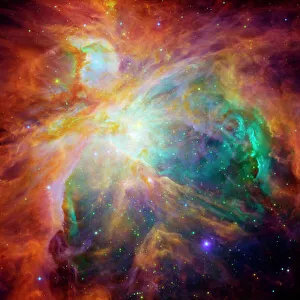
Embryonic wonders unfold in the depths of the Orion nebula, as captured in Picture No. 11675528


All Professionally Made to Order for Quick Shipping
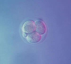
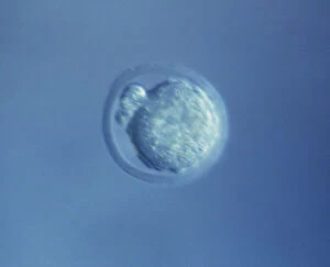
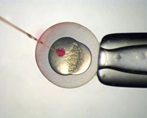
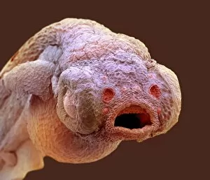
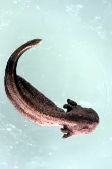
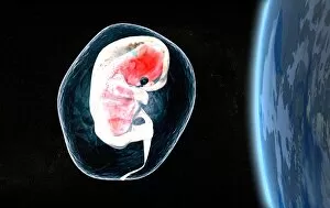
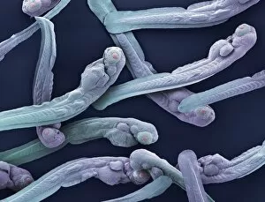
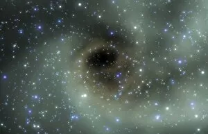
Embryonic wonders unfold in the depths of the Orion nebula, as captured in Picture No. 11675528. The cosmic nursery reveals a breathtaking display of celestial birth, where stars emerge from swirling clouds of gas and dust. In Picture No. 11675527, we witness a young star surrounded by a dusty protoplanetary disk, hinting at the potential for new planetary systems to form. But embryonic marvels are not limited to the vastness of space alone. Zooming into our own world, Picture No. 11675526 showcases zebrafish young under an SEM microscope – tiny embryos with intricate structures taking shape within their translucent bodies. Venturing further into uncharted territories, Explorer Sealife Giant Fish Corpse Pregnant Saw uncovers an astonishing sight: a colossal fish carrying its unborn offspring within its massive frame (Picture No. 12019792). This awe-inspiring example reminds us that even in nature's most unexpected corners, life begins anew. Delving deeper into the mysteries of creation, Picture No. 12479753 offers another glimpse into embryonic realms – this time featuring delicate marine organisms delicately suspended in fluid-filled sacs. And finally, returning to the grandeur of outer space once more, Picture No. 12479765 unveils yet another celestial spectacle: vibrant clusters of stars forming amidst glowing gas clouds like cosmic embryos on their journey towards illumination. In this captivating collection spanning both microcosms and macrocosms alike – from stellar nurseries to aquatic worlds – we are reminded that life's beginnings hold infinite beauty and promise.