
Electron Cryomicroscopy Collection
Electron cryomicroscopy (cryo-EM) has revolutionized the field of structural biology


All Professionally Made to Order for Quick Shipping
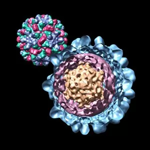
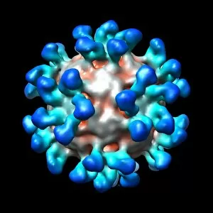
Electron cryomicroscopy (cryo-EM) has revolutionized the field of structural biology, allowing scientists to visualize intricate details of various viruses and their interactions with antibodies. Using this cutting-edge technique, researchers have captured stunning images of Bacteriophage phi29, Simian immunodeficiency virus (SIV), Cucumber necrosis virus, Human rhinovirus, and Ribgrass mosaic virus. These high-resolution computer models provide invaluable insights into the architecture and behavior of these viral entities. In one captivating image, a computer model showcases Bacteriophage phi29 in all its glory. This bacteriophage is known for its unique shape and ability to infect bacteria efficiently. The cryo-EM visualization allows us to appreciate the intricate structure that enables this tiny organism's remarkable functionality. Moving on to another fascinating subject, we delve into the realm of virology by exploring Simian immunodeficiency virus (SIV). Cryo-EM reveals the complex arrangement of proteins that make up SIV's outer shell. Understanding such structures aids in developing targeted therapies against similar viruses like HIV. The exploration continues as we encounter Cucumber necrosis virus through a detailed computer model generated using electron cryomicroscopy techniques. By deciphering its structure at an atomic level resolution, scientists gain crucial knowledge about how this plant pathogen operates within cucumber plants. Shifting our focus back to human health concerns, we investigate Human rhinovirus - one of the main culprits behind common colds. Through advanced imaging methods like cryo-EM combined with computational modeling approaches, researchers can study different strains and develop potential antiviral strategies. Additionally, electron cryomicroscopy provides us with a glimpse into how antibodies interact with Human rhinovirus particles. This visual representation helps elucidate mechanisms by which our immune system neutralizes viral threats effectively. Returning once again to bacteriophages – viruses that infect bacteria – we explore Bacteriophage P22.