Fraud Blocker Skip to main content

Ecto Parasite Collection (page 2)

"Exploring the Intricate World of Ecto Parasites: From Head Lice to Fleas and Beyond" Delving into the microscopic realm

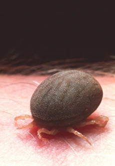
Background imageEcto Parasite Collection: Coloured SEM of a tick (Ixodes sp. ) in human skin

Coloured SEM of a tick (Ixodes sp. ) in human skin
Feeding tick. Coloured scanning electron micrograph (SEM) of a tick (Ixodes sp. ) feeding head- down in human skin. Ticks are arachnids which parasitise mammals, birds and reptiles

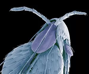
Background imageEcto Parasite Collection: Hedgehog flea, SEM

Hedgehog flea, SEM
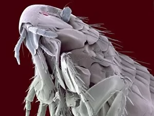
Hedgehog flea (Archaeopsylla erinacei), coloured scanning electron micrograph (SEM). The two structures protruding from the centre of the fleas head are sensory palps

Background imageEcto Parasite Collection: Female tick feeding on a human leg

Female tick feeding on a human leg

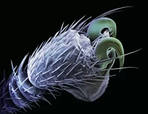
Background imageEcto Parasite Collection: Hedgehog flea claws, SEM

Hedgehog flea claws, SEM
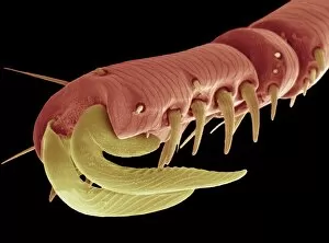
Hedgehog flea claws. Coloured scanning electron micrograph (SEM) of claws at the tip of a hedgehog fleas (Archaeopsylla erinacei) leg. The claws help to anchor the flea to its hosts fur

Background imageEcto Parasite Collection: Bat tick, SEM

Bat tick, SEM
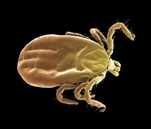
Bat tick. Coloured scanning electron micrograph (SEM) of a South American bat tick (order Acarina). Ticks are arachnid ectoparasites (external parasites)

Background imageEcto Parasite Collection: Relapsing fever tick

Relapsing fever tick. Macrophotograph of a relapsing fever tick (Ornithodorus moubata) feeding on human blood. This soft-bodied tick is a parasite of humans

Background imageEcto Parasite Collection: Squirrel flea, SEM

Squirrel flea, SEM
Squirrel flea (Orchopeas howardi howardi), coloured scanning electron micrograph (SEM). The two structures protruding from the centre of the fleas head are sensory palps

Background imageEcto Parasite Collection: Honey bee mite, SEM

Honey bee mite, SEM
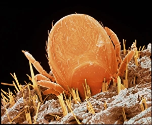
Honey bee mite. Coloured scanning electron micrograph (SEM) of a honey bee mite (Varroa sp.). Parasitic mites have decimated hives of wild and domesticated honey bees (Apis mellifera)

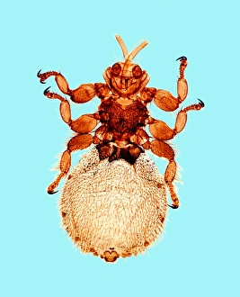
Background imageEcto Parasite Collection: Sheep ked

Sheep ked (Melophagus ovinus), light micrograph. This wingless, blood-sucking fly is an ectoparasite of sheep. The hooks on the ends of its legs help it to attach to its host

Background imageEcto Parasite Collection: Bat fly foot, SEM

Bat fly foot, SEM
Bat fly foot. Coloured scanning electron micrograph (SEM) of the foot of a bat fly (family Streblidae). These flies are ectoparasites (external parasites) of bats

Background imageEcto Parasite Collection: Bat fly head, SEM

Bat fly head, SEM
Bat fly head. Coloured scanning electron micrograph (SEM) of the head of a bat fly (family Streblidae). These flies are ectoparasites (external parasites) of bats

Background imageEcto Parasite Collection: Bat fly, SEM

Bat fly, SEM
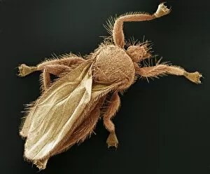
Bat fly. Coloured scanning electron micrograph (SEM) of a bat fly (family Streblidae). These flies are ectoparasites (external parasites) of bats

Background imageEcto Parasite Collection: Tick feeding, artwork

Tick feeding, artwork

Background imageEcto Parasite Collection: Eyelash mites, artwork

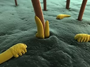
Eyelash mites, artwork

Background imageEcto Parasite Collection: Honeybee mite, artwork

Honeybee mite, artwork

Background imageEcto Parasite Collection: Eyelash mite, artwork

Eyelash mite, artwork

Background imageEcto Parasite Collection: Bedbug, artwork

Bedbug, artwork

Background imageEcto Parasite Collection: Cat flea, artwork

Cat flea, artwork

Background imageEcto Parasite Collection: Blood-filled tick, artwork

Blood-filled tick, artwork

Background imageEcto Parasite Collection: Head lice examination

Head lice examination
MODEL RELEASED. Head lice examination. Doctor checking a six year old girl for head lice (Pediculus humanus capitis). Head lice measure 2-3 millimetres in length

Background imageEcto Parasite Collection: Sarcoptic mange mite eggs, SEM

Sarcoptic mange mite eggs, SEM
Sarcoptic mange mites eggs, coloured scanning electron micrograph (SEM). This burrowing mite causes the skin disease scabies. Magnification: x200 when printed at 10 centimetres wide

Background imageEcto Parasite Collection: Sarcoptic mange mites, SEM

Sarcoptic mange mites, SEM
Sarcoptic mange mites, coloured scanning electron micrograph (SEM). These burrowing mites cause the skin disease scabies. Magnification: x30 when printed at 10 centimetres wide

Background imageEcto Parasite Collection: Monitor lizard tick

Monitor lizard tick (Amblyomma exornatum, formerly Aponomma exornatum). This tick is native to Africa, where, as the name suggests, it is mainly a parasite of monitor lizards

Background imageEcto Parasite Collection: Seal louse, SEM

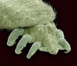
Seal louse, SEM
Seal louse (Echinophthirius horridus), coloured scanning electron micrograph (SEM). This ectoparasite (external parasite) feeds on the blood of seals, but only when the seals are on land

Background imageEcto Parasite Collection: Female lone star tick

Female lone star tick (Amblyomma americanum). This tick is found through the southeast and south-central United States. It is the vector for the spirochete bacteria Borrelia lonestari

Background imageEcto Parasite Collection: Male yellow dog tick

Male yellow dog tick (Amblyomma aureolatum). This tick is the vector for the bacteria Rickettsia rickettsii in Brazil. Rickettsia rickettsii, causes the often fatal Rocky Mountain spotted fever (RMSF)

Background imageEcto Parasite Collection: Trichodina parasite, SEM

Trichodina parasite, SEM
Trichodina parasite. Coloured scanning electron micrograph (SEM) of a Trichodina sp. protist on a freshwater minnow. This ciliate parasite lives on the skin


All Professionally Made to Order for Quick Shipping

"Exploring the Intricate World of Ecto Parasites: From Head Lice to Fleas and Beyond" Delving into the microscopic realm, we uncover a captivating world teeming with ecto parasites. Through scanning electron microscopy (SEM), we witness the minuscule yet formidable head louse, its tiny claws gripping onto human hair strands like a tightrope walker defying gravity. Moving on, our attention shifts to the sheep tick, another notorious ecto parasite that attaches itself firmly onto its host's skin. Under SEM, its intricate mouthparts resemble a menacing apparatus designed for bloodsucking survival. But not all ecto parasites evoke fear; some offer intriguing spectacles. Enter the flea circus, where these agile creatures perform acrobatic feats under magnification. Their vibrant colors and delicate structures astound as they leap through miniature hoops and balance on invisible tightropes. Venturing further into this hidden world reveals unexpected wonders. Eyelash mite tails come into focus under SEM, resembling wispy appendages that navigate their way amidst eyelashes without detection by their unsuspecting hosts. Intriguingly grotesque yet medically significant is the medicinal leech – an ancient remedy for various ailments. Its segmented body and powerful suction cups are revealed in vivid detail through SEM imagery. Stepping away from SEM but no less fascinating is the light micrograph showcasing Trichodina parasite - a circular organism with cilia adorning its surface like ethereal hairs dancing in water droplets. For those seeking unconventional curiosities within this realm of parasites, look no further than the colored SEM image capturing a flea penis in all its peculiar glory – an anatomical marvel tailored for reproduction. Zooming out slightly from individual organisms brings us to fleas mating captured under SEM - an intimate moment frozen in time as nature perpetuates itself through these tiny insects' union. Shifting gears slightly but still within this captivating ecosystem, we encounter the white-clawed freshwater crayfish.