Fraud Blocker Skip to main content

Diagnostic Collection (page 3)

"Unlocking the Secrets of Health: Exploring the World Tools" Medieval Urine Wheel: From ancient times to modern medicine

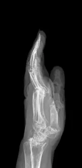
Background imageDiagnostic Collection: Arthritic hand, X-ray C017 / 7171

Arthritic hand, X-ray C017 / 7171
Arthritic hand. X-ray of the hand and wrist of a 56 year old patient with rheumatoid arthritis. Rheumatoid arthritis is caused by the immune system attacking the bodys own tissues

Background imageDiagnostic Collection: Secondary bone cancer, X-ray C017 / 7162

Secondary bone cancer, X-ray C017 / 7162
Secondary bone cancer. X-ray of the spine of a 40 year old female with metastatic (secondary) bone cancer (bright white areas). The cancer has metastasised (spread) from a primary cancer

Background imageDiagnostic Collection: Hicks patent thermometer, circa 1880

Hicks patent thermometer, circa 1880
Hicks patent medical thermometer. James Joseph Hicks (1837-1916), was a British manufacturer of scientific instruments who filed several patents of medical and meteorological thermometers

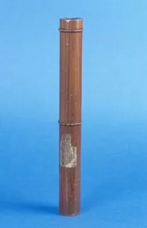
Background imageDiagnostic Collection: Laennec monaural stethoscope, circa 1820 C017 / 6951

Laennec monaural stethoscope, circa 1820 C017 / 6951
English monaural stethoscope, made out of fruitwood, dating from the year 1820. French physician, Rene Theophile Hyacinthe Laennec invented the monaural stethoscope in 1816

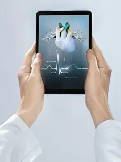
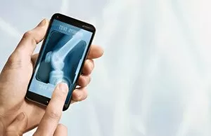
Background imageDiagnostic Collection: Telemedicine, conceptual image C017 / 7605

Telemedicine, conceptual image C017 / 7605
MODEL RELEASED. Telemedicine, conceptual image

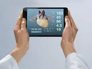
Background imageDiagnostic Collection: Telemedicine, conceptual image C017 / 7592

Telemedicine, conceptual image C017 / 7592
MODEL RELEASED. Telemedicine, conceptual image

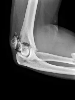
Background imageDiagnostic Collection: Broken elbow, X-ray C017 / 7583

Broken elbow, X-ray C017 / 7583
Broken elbow. X-ray of the arm of a 55 year old male patient with fractures to the elbow and radius (upper lower arm bone)

Background imageDiagnostic Collection: Telemedicine, conceptual image C017 / 7590

Telemedicine, conceptual image C017 / 7590
Telemedicine, conceptual image

Background imageDiagnostic Collection: Telemedicine, conceptual image C017 / 7607

Telemedicine, conceptual image C017 / 7607
Telemedicine, conceptual image

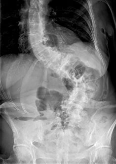
Background imageDiagnostic Collection: Scoliosis of the spine, X-ray C017 / 7167

Scoliosis of the spine, X-ray C017 / 7167
Scoliosis. X-ray of the spine of a 71 year old female patient with severe scoliosis (sideways curvature). This condition usually starts in infancy or adolescence, and is most often seen in girls

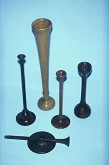
Background imageDiagnostic Collection: Monaural stethoscopes, 19th century C017 / 6931

Monaural stethoscopes, 19th century C017 / 6931
Five monaural stethoscopes dating from between 1860 and 1890. These are made from ebony and fruitwood. French physician, Rene Theophile Hyacinthe Laennec invented the monaural stethoscope in 1816

Background imageDiagnostic Collection: Phonendoscope, circa 1900 C017 / 6955

Phonendoscope, circa 1900 C017 / 6955
Phonendoscope dating from around 1900, England. This device was used in the early 20th century for listening to internal sounds of the body (auscultation)

Background imageDiagnostic Collection: Telemedicine, conceptual image C017 / 7601

Telemedicine, conceptual image C017 / 7601
Telemedicine, conceptual image

Background imageDiagnostic Collection: Extra long monaural stethoscope, circa 18 C017 / 6948

Extra long monaural stethoscope, circa 18 C017 / 6948
Unusual extra long monaural stethoscope dating from 1860. This piece was made in England out of fruitwood. French physician

Background imageDiagnostic Collection: Telemedicine, conceptual image C017 / 7603

Telemedicine, conceptual image C017 / 7603
MODEL RELEASED. Telemedicine, conceptual image

Background imageDiagnostic Collection: Oriental figure for diagnosis, circa 1830 C017 / 0738

Oriental figure for diagnosis, circa 1830 C017 / 0738
Recumbent figure for diagnosis, carved from ivory on a wooden base. This object is Chinese and dates from around 1830

Background imageDiagnostic Collection: Telemedicine, conceptual image C017 / 7599

Telemedicine, conceptual image C017 / 7599
Telemedicine, conceptual image

Background imageDiagnostic Collection: Lung disease, X-ray C017 / 7568

Lung disease, X-ray C017 / 7568
Lung disease. X-ray of the chest of a 88 year old male with a history of lung disease. The patient has previously had part of the right lung (seen at left) removed, a lobectomy

Background imageDiagnostic Collection: Bennet percussor, circa 1860 C017 / 6934

Bennet percussor, circa 1860 C017 / 6934
Bennet percussor with ivory pleximeter dating from 1860s, England. A percussor is a small hammer used for testing reflexes and for percussive examinations of the chest

Background imageDiagnostic Collection: Telemedicine, conceptual image C017 / 7594

Telemedicine, conceptual image C017 / 7594
MODEL RELEASED. Telemedicine, conceptual image

Background imageDiagnostic Collection: Bennet percussor, circa 1860 C017 / 6971

Bennet percussor, circa 1860 C017 / 6971
Bennet percussor dating from 1860s, England. A percussor is a small hammer used for testing reflexes and for percussive examinations of the chest

Background imageDiagnostic Collection: Contraceptive implant, X-ray C017 / 7390

Contraceptive implant, X-ray C017 / 7390
Contraceptive implant. X-ray of the arm of a 19 year old female showing a contraceptive implant (thin rod at upper centre)

Background imageDiagnostic Collection: Telemedicine, conceptual image C017 / 7606

Telemedicine, conceptual image C017 / 7606
Telemedicine, conceptual image

Background imageDiagnostic Collection: Polycystic kidneys, ultrasound scan

Polycystic kidneys, ultrasound scan
Polycystic kidneys. Ultrasound scan of the kidney of an 82 year old male showing numerous fluid-filled cysts (dark areas)

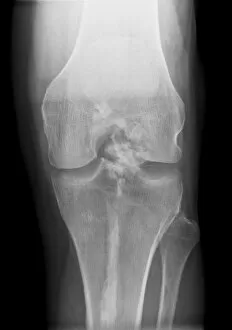
Background imageDiagnostic Collection: Melorheostosis of the knee, X-ray C017 / 7146

Melorheostosis of the knee, X-ray C017 / 7146
Melorheostosis of the knee. X-ray of left knee of a 52 year old male patient showing damage (white area at centre) caused by melorheostosis

Background imageDiagnostic Collection: Medical care, conceptual artwork F006 / 3817

Medical care, conceptual artwork F006 / 3817
Medical care, conceptual computer artwork

Background imageDiagnostic Collection: Home care, conceptual artwork F006 / 3859

Home care, conceptual artwork F006 / 3859
Home care, conceptual computer artwork

Background imageDiagnostic Collection: Medical care, conceptual artwork F006 / 3979

Medical care, conceptual artwork F006 / 3979
Medical care, conceptual computer artwork

Background imageDiagnostic Collection: Heart health, conceptual artwork F006 / 3776

Heart health, conceptual artwork F006 / 3776
Heart health, conceptual computer artwork

Background imageDiagnostic Collection: Online diagnosis, conceptual artwork F006 / 3899

Online diagnosis, conceptual artwork F006 / 3899
Online diagnosis, conceptual computer artwork

Background imageDiagnostic Collection: Blood sample tube F006 / 7249

Blood sample tube F006 / 7249
Blood sample tube

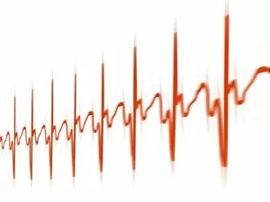
Background imageDiagnostic Collection: ECG, artwork F006 / 4606

ECG, artwork F006 / 4606
ECG. Computer artwork of an electrocardiogram (ECG) showing a normal heart rate

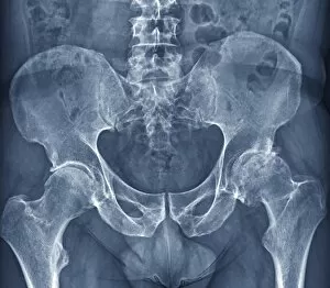
Background imageDiagnostic Collection: Osteoarthritis of the hip, X-ray F006 / 3744

Osteoarthritis of the hip, X-ray F006 / 3744
Osteoarthritis of the hip. Coloured frontal X-ray (front view) of the pelvis in a 59 year old patient showing osteoarthritis of both hip joints

Background imageDiagnostic Collection: Online diagnosis, conceptual artwork F006 / 3892

Online diagnosis, conceptual artwork F006 / 3892
Online diagnosis, conceptual computer artwork

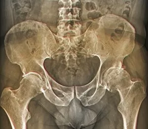
Background imageDiagnostic Collection: Osteoarthritis of the hip, X-ray F006 / 3745

Osteoarthritis of the hip, X-ray F006 / 3745
Osteoarthritis of the hip. Coloured frontal X-ray (front view) of the pelvis in a 59 year old patient showing osteoarthritis of both hip joints

Background imageDiagnostic Collection: Heart monitoring, conceptual image F008 / 3202

Heart monitoring, conceptual image F008 / 3202
Heart monitoring, conceptual image

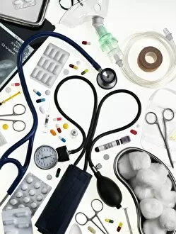
Background imageDiagnostic Collection: Medical equipment and drugs F008 / 3189

Medical equipment and drugs F008 / 3189
Medical equipment and drugs

Background imageDiagnostic Collection: Medical equipment and drugs F008 / 3190

Medical equipment and drugs F008 / 3190
Medical equipment and drugs

Background imageDiagnostic Collection: Blood pressure pills, conceptual image F008 / 3174

Blood pressure pills, conceptual image F008 / 3174
Blood pressure pills, conceptual image

Background imageDiagnostic Collection: Medical equipment and drugs F008 / 3188

Medical equipment and drugs F008 / 3188
Medical equipment and drugs

Background imageDiagnostic Collection: Heart monitoring, conceptual image F008 / 3204

Heart monitoring, conceptual image F008 / 3204
Heart monitoring, conceptual image

Background imageDiagnostic Collection: ECG, computer artwork

ECG, computer artwork
ECG. Computer artwork of an electrocardiogram (ECG) showing a normal heart rate

Background imageDiagnostic Collection: Stethoscope F005 / 0841

Stethoscope F005 / 0841
Stethoscope

Background imageDiagnostic Collection: Blood pressure gauge F005 / 0844

Blood pressure gauge F005 / 0844
Blood pressure gauge

Background imageDiagnostic Collection: Capsule endoscope, artwork F005 / 0186

Capsule endoscope, artwork F005 / 0186
Capsule endoscope, computer artwork

Background imageDiagnostic Collection: Blood pressure gauge F005 / 0843

Blood pressure gauge F005 / 0843
Blood pressure gauge

Background imageDiagnostic Collection: Blood pressure gauge F005 / 0842

Blood pressure gauge F005 / 0842
Blood pressure gauge

Background imageDiagnostic Collection: Medical test tube with blood, artwork

Medical test tube with blood, artwork
Computer artwork of a medical test tube with blood samples and a DNA helix, depicting dna profiling, genetic testing and other DNA based tests


All Professionally Made to Order for Quick Shipping

"Unlocking the Secrets of Health: Exploring the World Tools" Medieval Urine Wheel: From ancient times to modern medicine, urine analysis has been a diagnostic tool used to detect various health conditions. Eyesight Test Chart: A simple yet crucial diagnostic tool that helps determine visual acuity and identify potential eye problems. Broken Wrist Bone, X-ray C017 / 7187: X-rays provide a clear view of fractured bones, aiding in accurate diagnosis and guiding treatment plans. Panoramic Dental X-ray: This comprehensive imaging technique reveals dental issues like cavities, impacted teeth, or jaw abnormalities for effective treatment planning. Home Cholesterol Test Kit: Empowering individuals with easy-to-use kits to monitor cholesterol levels at home and take proactive steps towards heart health. Full Body Scan, MRI Scan: Non-invasive and highly detailed imaging technology that allows doctors to visualize internal organs and diagnose complex medical conditions accurately. Brain Anatomy, MRI Scan: Advanced brain imaging enables neurologists to study intricate structures within the brain for precise diagnoses of neurological disorders. ECGs of a Normal Heart Rate, Artwork: Electrocardiograms help assess heart function by recording electrical activity; this artwork showcases what a healthy heartbeat looks like on paper. Tuberculosis, X-ray: Chest x-rays play a vital role in diagnosing tuberculosis by detecting characteristic lung abnormalities associated with this infectious disease. Normal EEG Readout of the Brain's Alpha Waves: Electroencephalography records brain wave patterns helping neurologists evaluate brain function and diagnose epilepsy or sleep disorders effectively. Eye Examination:A thorough examination assessing vision sharpness (visual acuity), peripheral vision tests (visual field), color perception evaluation among others is essential for maintaining good eye health Fractured Ankle, X-Ray.