Fraud Blocker Skip to main content

Diagnosing Collection (page 2)

"Unveiling the Art of Diagnosing: From Ancient Practices to Modern Techniques" In this captivating image, a young boy can be seen measuring his temperature

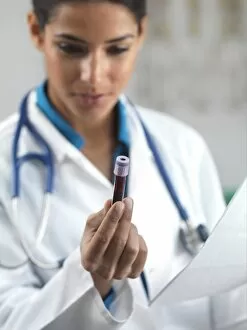
Background imageDiagnosing Collection: Doctor holding a blood sample C013 / 8842

Doctor holding a blood sample C013 / 8842
Doctor holding a blood sample

Background imageDiagnosing Collection: Paediatric examination C013 / 8840

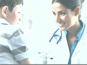
Paediatric examination C013 / 8840
Paediatric examination. Doctor talking to a 7-year-old boy
Background imageDiagnosing Collection: Doctor holding a blood sample C013 / 8841

Doctor holding a blood sample C013 / 8841
Doctor holding a blood sample

Background imageDiagnosing Collection: Paediatric examination C013 / 8838

Paediatric examination C013 / 8838
Paediatric examination. Doctor talking to a 7-year-old boy

Background imageDiagnosing Collection: Paediatric examination C013 / 8839

Paediatric examination C013 / 8839
Paediatric examination. Doctor talking to a 7-year-old boy

Background imageDiagnosing Collection: Paediatric examination C013 / 8837

Paediatric examination C013 / 8837
Paediatric examination. Doctor talking to a 7-year-old boy

Background imageDiagnosing Collection: Paediatric examination C013 / 8833

Paediatric examination C013 / 8833
Paediatric examination. Doctor checking a 7-year-old boys glands to see if they are swollen. Swelling of the glands (lymphadenopathy)

Background imageDiagnosing Collection: Paediatric examination C013 / 8832

Paediatric examination C013 / 8832
Paediatric examination. Doctor checking a 7-year-old boys glands to see if they are swollen. Swelling of the glands (lymphadenopathy)

Background imageDiagnosing Collection: Paediatric examination C013 / 8829

Paediatric examination C013 / 8829
Paediatric examination. Doctor talking to a 7-year-old boy

Background imageDiagnosing Collection: Paediatric examination C013 / 8831

Paediatric examination C013 / 8831
Paediatric examination. Doctor checking a 7-year-old boys glands to see if they are swollen. Swelling of the glands (lymphadenopathy)

Background imageDiagnosing Collection: Paediatric examination C013 / 8830

Paediatric examination C013 / 8830
Paediatric examination. Doctor checking a 7-year-old boys glands to see if they are swollen. Swelling of the glands (lymphadenopathy)

Background imageDiagnosing Collection: Paediatric examination C013 / 8828

Paediatric examination C013 / 8828
Paediatric examination. Doctor cleaning a wound on the arm of a 3-year-old girl

Background imageDiagnosing Collection: Paediatric examination C013 / 8825

Paediatric examination C013 / 8825
Paediatric examination. Doctor checking a 3-year-old boys glands to see if they are swollen. Swelling of the glands (lymphadenopathy)

Background imageDiagnosing Collection: Paediatric examination C013 / 8827

Paediatric examination C013 / 8827
Paediatric examination. Doctor talking to a 3-year-old girl

Background imageDiagnosing Collection: Paediatric examination C013 / 8824

Paediatric examination C013 / 8824
Paediatric examination. Doctor using a stethoscope to listen to a 3-year-old boys lungs. A stethoscope is used to listen to sounds within the body

Background imageDiagnosing Collection: Paediatric examination C013 / 8826

Paediatric examination C013 / 8826
Paediatric examination. Doctor talking to a 3-year-old girl

Background imageDiagnosing Collection: Irregular heart beat, conceptual image C013 / 8807

Irregular heart beat, conceptual image C013 / 8807
Irregular heart beat, conceptual image. Computer artwork of ECG (electrocardiogram) traces (red) behind a human heart, representing cardiac arrhythmia

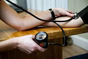
Background imageDiagnosing Collection: Blood pressure measurement C013 / 7569

Blood pressure measurement C013 / 7569
Blood pressure measurement. Patient having their blood pressure taken at a doctors surgery

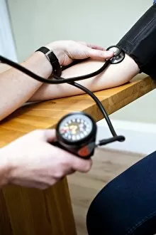
Background imageDiagnosing Collection: Blood pressure measurement C013 / 7568

Blood pressure measurement C013 / 7568
Blood pressure measurement. Patient having their blood pressure taken at a doctors surgery

Background imageDiagnosing Collection: Automatic DNA analyser, computer artwork

Automatic DNA analyser, computer artwork
Instant DNA analysis. Computer artwork of a machine that can instantly analyse DNA samples to warn of genetic diseases

Background imageDiagnosing Collection: Dental X-ray

Dental X-ray
MODEL RELEASED. Dental X-ray. Dentist looking at an X-ray of a patients teeth. This is a panoramic wraparound X-ray that shows all the teeth of both the upper and lower jaw

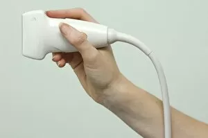
Background imageDiagnosing Collection: Ultrasound transducer

Ultrasound transducer
MODEL RELEASED. Ultrasound transducer. This device emits ultrasonic waves and detects the reflected echoes. The change in frequency is used to build images of the inside of the body

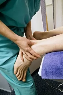
Background imageDiagnosing Collection: Podiatric ultrasound

Podiatric ultrasound
MODEL RELEASED. Podiatric ultrasound. Podiatrist performing an ultrasound examination on the sole of a foot. This technique is used to diagnose soft tissue problems in the foot

Background imageDiagnosing Collection: Studying the brain, conceptual image

Studying the brain, conceptual image

Background imageDiagnosing Collection: Pulse measurement, 14th century artwork

Pulse measurement, 14th century artwork
Pulse measurement. 14th century artwork of a doctor measuring a patients pulse. As seen here, the pulse can be taken at the wrist or in the armpit

Background imageDiagnosing Collection: Patient examination, 14th century artwork

Patient examination, 14th century artwork
Patient examination. 14th century artwork of a doctor examining a patient by palpation of the abdomen. Artwork from Anathomia (1345), by the Italian anatomist Guido da Vigevano

Background imageDiagnosing Collection: Hip joint examination

Hip joint examination
MODEL RELEASED. Hip joint examination. Physiotherapist performing an assessment of the hip joints of a young boy. The bones of the hip are held together by ligaments

Background imageDiagnosing Collection: Knee reflex test

Knee reflex test
MODEL RELEASED. Knee reflex test. Physiotherapist using a testing mallet to perform a knee reflex test on a young child. The mallet is used to gently tap a specific area just below the childs knee

Background imageDiagnosing Collection: Cervical spine examination

Cervical spine examination
MODEL RELEASED. Cervical spine examination. Physiotherapist manipulating the cervical (neck) region of a young boys spine, during a spinal examination

Background imageDiagnosing Collection: Foot and ankle examination

Foot and ankle examination
MODEL RELEASED. Foot and ankle examination. Physiotherapist preparing to perform a foot and ankle examination on a young child

Background imageDiagnosing Collection: Shoulder examination

Shoulder examination
MODEL RELEASED. Shoulder examination. Physiotherapist manipulating a patients arm during an examination of his shoulder joint. This is known as a glenohumeral examination

Background imageDiagnosing Collection: Achillies tendon examination

Achillies tendon examination
MODEL RELEASED. Achillies tendon examination. Physiotherapist using her hands to examine and treat a patients Achilles tendon

Background imageDiagnosing Collection: Apleys knee test

Apleys knee test
MODEL RELEASED. Apleys knee test. Physiotherapist performing an Apleys knee test to assess a patients knee injury. This test is carried out to diagnose knee cartilage injuries

Background imageDiagnosing Collection: Lachman knee ligament test

Lachman knee ligament test
MODEL RELEASED. Lachman knee ligament test. Physiotherapist performing a Lachman test on a patients knee ligaments. This test is designed as an assessment of the knees anterior cruciate ligament

Background imageDiagnosing Collection: McMurrays knee joint test

McMurrays knee joint test
MODEL RELEASED. McMurrays knee joint test. Physiotherapist performing a McMurrays knee joint test on a patient. The McMurray test is designed to assess injuries to the cartilage in the knee

Background imageDiagnosing Collection: X-ray diagnosis

X-ray diagnosis
MODEL RELEASED. X-ray diagnosis. Doctor examining an X-ray of a hand

Background imageDiagnosing Collection: Radiologist studying an X-ray

Radiologist studying an X-ray
MODEL RELEASED. Radiologist studying an X-ray

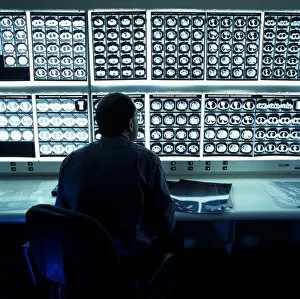
Background imageDiagnosing Collection: Studying CT scans

Studying CT scans
MODEL RELEASED. Studying CT scan. Doctor studying computer tomography (CT) scans of horizontal sections through a patients chest and abdomen

Background imageDiagnosing Collection: Doctor holding MRI scans

Doctor holding MRI scans
MODEL RELEASED. Doctor holding MRI scans. The scans are sagittal (vertical and sideways) sections through a patients head

Background imageDiagnosing Collection: Doctor examining MRI scans

Doctor examining MRI scans
MODEL RELEASED. Doctor examining MRI scans. The scans are sagittal (vertical and sideways) sections through a patients head

Background imageDiagnosing Collection: Radiologists examining an X-ray

Radiologists examining an X-ray
MODEL RELEASED. Radiologists studying an X-ray of a patients chest

Background imageDiagnosing Collection: Doctors examining MRI scans

Doctors examining MRI scans
MODEL RELEASED. Doctors examining MRI scans. The scans are sagittal (side view) sections through a patients head. MRI is an important diagnostic tool that allows imaging of internal structures

Background imageDiagnosing Collection: Blood pressure measurement

Blood pressure measurement

Background imageDiagnosing Collection: Wrist pulse, 17th century

Wrist pulse, 17th century
Wrist pulse. Artwork demonstrating how to take the pulse in the wrist, as depicted by the Chinese. This artwork was published in Europe in Specimen Medicinae Sinicae (1682)

Background imageDiagnosing Collection: Tuberculosis diagnosis, 19th century

Tuberculosis diagnosis, 19th century. Artwork showing the French physician Rene Laennec (1781-1826), at the Necker Hospital, Paris, France, examining a patient (left)

Background imageDiagnosing Collection: Bone density scanner, composite image

Bone density scanner, composite image
MODEL RELEASED. Bone density scanner. Composite image of a dual energy X-ray absorptiometry (DXA) scanner being used to measure a patients bone mineral density (BMD)

Background imageDiagnosing Collection: Ultrasound DVT scanning, composite image

Ultrasound DVT scanning, composite image
MODEL RELEASED. Ultrasound DVT scanning. Composite image of a radiologists hand using an ultrasound transducer to scan a patients leg for a deep vein thrombosis (DVT)

Background imageDiagnosing Collection: Chest X-ray imaging, composite image

Chest X-ray imaging, composite image. At left a ceiling-mounted X-ray machine is being used to X-ray a patients chest. The artwork of the patient (at right)


All Professionally Made to Order for Quick Shipping

"Unveiling the Art of Diagnosing: From Ancient Practices to Modern Techniques" In this captivating image, a young boy can be seen measuring his temperature, symbolizing the timeless pursuit illnesses. The journey of diagnosis dates back centuries, with pioneers like Abulcasis, an Islamic physician who laid the foundation for medical knowledge. The illustration from "The Works of Hippocrates" showcases an oral examination, highlighting how ancient physicians meticulously observed symptoms and signs to unravel mysteries hidden within the human body. Even then, patients would tremble at the hand of doctors as depicted in another intriguing illustration. Did you know that George Washington himself turned into a physician? This engraving reminds us that even great leaders recognized the importance of understanding ailments and providing care. Richard Morton's contribution cannot be overlooked either; he made significant strides in identifying diseases such as tuberculosis. Dental plaque and tartar are also part of this diagnostic puzzle - their presence revealing more than just dental health but potential systemic issues too. Fast forward to modern times where radiologists examine knee MRI scans with precision (C014 / 1300). X-rays have become invaluable tools in detecting throat cancer (X-rays) while Oriental figures from circa 1830 remind us that different cultures had unique approaches to diagnosis (C017 / 0738). Medical alchemists from the 18th century sought answers through experimentation and exploration. Their practices paved the way for advancements we benefit from today. Lastly, a urine diabetes test (C014 / 1231) serves as a reminder that diagnostics extend beyond visual observations – it involves analyzing bodily fluids too. From ancient wisdom to cutting-edge technology, it has evolved immensely over time, and is through these collective efforts that we continue to unlock medical mysteries and provide better care for all.