Fraud Blocker Skip to main content

Dermatological Collection

"Dermatological Wonders: Exploring the Artistry of Skin Disorders" Delve into the intricate world of dermatology, where skin disorders become captivating works of art

Background imageDermatological Collection: Skin disorders, artwork

Skin disorders, artwork
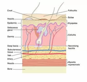
Skin disorders. Computer artwork showing the structure of the skin, and the layers affected by various skin disorders. Folliculitis is the inflammation of hair follicles; Bullae are blisters;

Background imageDermatological Collection: Human skin, illustration

Human skin, illustration
3d illustration of a cross section of a hair follicle with description

Background imageDermatological Collection: Dermatoscope

Dermatoscope. This instrument is used to examine the skin. It includes a magnifier and a light source

Background imageDermatological Collection: Hypo-allergenic soap

Hypo-allergenic soap. Top view of a bar of soap that has been developed so as not to cause an allergic reaction when used

Background imageDermatological Collection: Skin surface, SEM

Skin surface, SEM
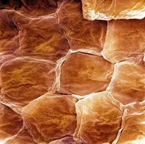
Skin surface. Coloured scanning electron micrograph (SEM) of squamous cells on the surface of the skin. These are flat, keratinised

Background imageDermatological Collection: Favus, from a book by Daniel Cornelius Danielssen (1815-94) 1892 (coloured engraving)

Favus, from a book by Daniel Cornelius Danielssen (1815-94) 1892 (coloured engraving)
CHT236758 Favus, from a book by Daniel Cornelius Danielssen (1815-94) 1892 (coloured engraving) by Norwegian School, (19th century); Private Collection; eArchives Charmet; Norwegian, out of copyright

Background imageDermatological Collection: Skin disorders, c. 1890 (colour chromolithograph)

Skin disorders, c. 1890 (colour chromolithograph)
1065620 Skin disorders, c.1890 (colour chromolithograph) by German School, (19th century); Private Collection; Prismatic Pictures; German, out of copyright

Background imageDermatological Collection: Advertisement for Viola Cream, c. 1890 (colour litho)

Advertisement for Viola Cream, c. 1890 (colour litho)
HGL396896 Advertisement for Viola Cream, c.1890 (colour litho) by American School, (19th century); Hagley Museum & Library, Wilmington, Delaware

Background imageDermatological Collection: Advertisement for Dr. Hebras Viola Cream, c. 1897 (colour litho)

Advertisement for Dr. Hebras Viola Cream, c. 1897 (colour litho)
HGL396904 Advertisement for Dr. Hebras Viola Cream, c.1897 (colour litho) by American School, (19th century); Hagley Museum & Library, Wilmington, Delaware

Background imageDermatological Collection: Congenital Ichthyosis, from a book by G. M. Olbers, 1830 (coloured engraving)

Congenital Ichthyosis, from a book by G. M. Olbers, 1830 (coloured engraving)
CHT236760 Congenital Ichthyosis, from a book by G.M. Olbers, 1830 (coloured engraving) by French School, (19th century); Bibliotheque de l Institut d Ophtalmologie, Paris

Background imageDermatological Collection: Ringworm, from a book by Baron Jean Louis Alibert (1768-1837) 1838 (coloured engraving)

Ringworm, from a book by Baron Jean Louis Alibert (1768-1837) 1838 (coloured engraving)
CHT236757 Ringworm, from a book by Baron Jean Louis Alibert (1768-1837) 1838 (coloured engraving) by French School, (19th century); Bibliotheque de la Faculte de Medecine, Paris

Background imageDermatological Collection: Yeast fungus skin infection, SEM

Yeast fungus skin infection, SEM
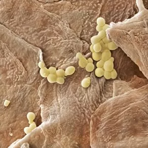
Yeast fungus skin infection. Coloured scanning electron micrograph (SEM) of human skin infected with a yeast fungus. Magnification: x3130 when printed at 10 centimetres wide

Background imageDermatological Collection: Psoriasis

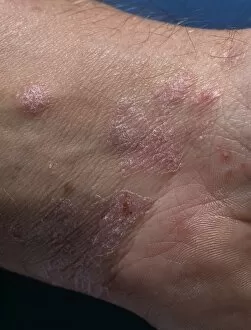
Psoriasis lesions on the skin of a mans wrist. Psoriasis is a common skin disease involving a large increase in the speed of epidermal (outer skin layer) cell production

Background imageDermatological Collection: Propionibacterium acnes C014 / 0631

Propionibacterium acnes C014 / 0631
Propionibacterium acnes bacteria, coloured scanning electron micrograph (SEM). This bacteria is found deep in the hair follicles and pores of the skin

Background imageDermatological Collection: Pigmented skin mole C013 / 7318

Pigmented skin mole C013 / 7318
MODEL RELEASED. Pigmented skin mole. Pigmented naevus or mole on the skin of a 24-year-old man. Also known as a melanocytic, or melanotic, naevus

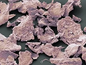
Background imageDermatological Collection: Flakes of dead skin, SEM

Flakes of dead skin, SEM
Flakes of dead skin. Coloured scanning electron micrograph (SEM) of dead skin from the scalp (dandruff), a condition where there is an excessive shedding of dead skin cells

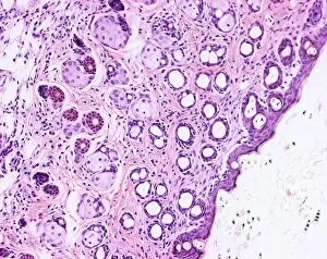
Background imageDermatological Collection: Skin and hair follicles, light micrograph

Skin and hair follicles, light micrograph
Skin and hair follicles. Light micrograph of a section through skin, showing hair follicles (dark purple circular structures). Haematoxylin and eosin stain

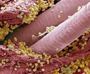
Background imageDermatological Collection: Lymphocytes in hair follicle, SEM

Lymphocytes in hair follicle, SEM
Lymphocytes in hair follicle, coloured scanning electron micrograph (SEM). Large numbers of lymphocytes (yellow) are seen in inflamed and necrotic tissue at the base of hair shafts (red)

Background imageDermatological Collection: Elderly womans hands

Elderly womans hands
MODEL RELEASED. Elderly womans hands. She is 93 years old

Background imageDermatological Collection: Cutaneous leishmaniasis, 1917

Cutaneous leishmaniasis, 1917
Cutaneous leishmaniasis. Lesions on the face of a man in Palestine in 1917 in a case of cutaneous leishmaniasis. The local name for this condition at the time was Jericho buttons

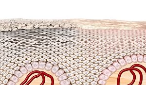
Background imageDermatological Collection: Skin anatomy, artwork

Skin anatomy, artwork
Skin anatomy. Artwork of a section through human skin, showing the flattening of skin cells as they rise to the dead keratinised surface layer (top) from the living layer below (the dermis)

Background imageDermatological Collection: Hair follicle, illustration C018 / 0795

Hair follicle, illustration C018 / 0795
Hair follicle, illustration. Hair is formed from the protein keratin. It is a dead tissue that is pushed up by growth from the bulb at the base of the follicle

Background imageDermatological Collection: Ferdinand von Hebra, Austrian physician C018 / 7109

Ferdinand von Hebra, Austrian physician C018 / 7109
Ferdinand von Hebra (1816-1880). 1889 engraving of the Austrian physician and dermatologist Ferdinand Ritter von Hebra. Von Hebra is regarded as the founder of the scientific theory of skin diseases

Background imageDermatological Collection: Plegmon eye inflammation, artwork

Plegmon eye inflammation, artwork
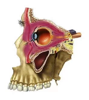
Plegmon eye inflammation. Artwork of a sagittal section through a human eye and the surrounding structures, in a case of a subcutaneous (below the skin) phlegmon inflammation of an eye cavity

Background imageDermatological Collection: Sweat gland in skin, artwork

Sweat gland in skin, artwork
Sweat gland in skin. Artwork of a section of human skin (top) with the upper skin layers shown, along with the duct leading from a sweat pore to its sweat gland (bottom)

Background imageDermatological Collection: Wart structure, artwork

Wart structure, artwork
Wart structure. Artwork of a section through human skin, showing the subsurface blood vessels (red) supplying a wart. Warts are harmless

Background imageDermatological Collection: Wart, artwork

Wart, artwork
Wart. Computer artwork of a wart (Verruca vulgaris) forming on the surface of the skin. Warts are harmless, contagious growths of the outermost layer of the skin (epidermis)

Background imageDermatological Collection: Propionibacterium acnes C014 / 0635

Propionibacterium acnes C014 / 0635
Propionibacterium acnes bacteria, coloured scanning electron micrograph (SEM). This bacteria is found deep in the hair follicles and pores of the skin

Background imageDermatological Collection: Propionibacterium acnes C014 / 0634

Propionibacterium acnes C014 / 0634
Propionibacterium acnes bacteria, coloured scanning electron micrograph (SEM). This bacteria is found deep in the hair follicles and pores of the skin

Background imageDermatological Collection: Propionibacterium acnes C014 / 0633

Propionibacterium acnes C014 / 0633
Propionibacterium acnes bacteria, coloured scanning electron micrograph (SEM). This bacteria is found deep in the hair follicles and pores of the skin

Background imageDermatological Collection: Propionibacterium acnes C014 / 0630

Propionibacterium acnes C014 / 0630
Propionibacterium acnes

Background imageDermatological Collection: Skin surface, SEM C017 / 8494

Skin surface, SEM C017 / 8494
Skin surface. Coloured scanning electron micrograph (SEM) of vapour entrapments/sweat globules on the surface of human skin

Background imageDermatological Collection: Skin surface, SEM C017 / 8493

Skin surface, SEM C017 / 8493
Skin surface. Coloured scanning electron micrograph (SEM) of vapour entrapments/sweat globules on the surface of human skin

Background imageDermatological Collection: Ultraviolet light animal tests, 1926 C017 / 7867

Ultraviolet light animal tests, 1926 C017 / 7867
Ultraviolet light animal tests. Researchers for the United States Department of Agriculture carrying out ultraviolet (UV) light therapy on white rats

Background imageDermatological Collection: Ointment C016 / 4687

Ointment C016 / 4687
Ointment being squeezed from a tube

Background imageDermatological Collection: Hospital phototherapy session, 1920s C016 / 2384

Hospital phototherapy session, 1920s C016 / 2384
Hospital phototherapy. Physiotherapy nurse positioning an ultraviolet (UV) Alpine Sun Lamp over a patients hand during a session of phototherapy

Background imageDermatological Collection: Carpet beetle hair, SEM

Carpet beetle hair, SEM
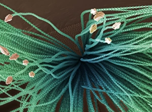
Carpet beetle hair. Coloured scanning electron micrograph (SEM) of the spear-headed hair (hastisetae) of a carpet beetle larva (family Dermestidae)

Background imageDermatological Collection: Corneal strip

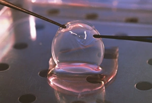
Corneal strip grown in the laboratory. It has been cultured from human epithelial cells that line the cornea, the outer shell of the eyeball

Background imageDermatological Collection: Second-degree burn, artwork

Second-degree burn, artwork
Second-degree burn, cut-away artwork. A second-degree burn damages the top layer of skin (epidermis, dark pink) and the underlying tissue (dermis, pink)

Background imageDermatological Collection: Third-degree burn, artwork

Third-degree burn, artwork
Third-degree burn, cut-away artwork. A third-degree burn is a severe burn that damages the top layer of skin (epidermis, dark pink), the underlying tissue (dermis)

Background imageDermatological Collection: Eczema skin condition

Eczema skin condition
Eczema. Computer artwork of the epidermis (upper skin layer) of skin affected by eczema. Eczema is a skin inflammation characterised by an itchy pink rash on the skins surface

Background imageDermatological Collection: Eczema, SEM

Eczema, SEM
Eczema, coloured scanning electron micrograph (SEM). Eczema is a form of dermatitis or inflammation of the upper layers of the skin

Background imageDermatological Collection: Psoriasis plaques on a hand

Psoriasis plaques on a hand
Psoriasis on the hand. Artwork from Robert Willans 1808 medical textbook, On Cutaneous Diseases, showing what he calls psoriasis palmaria, or psorasis affecting the palm of the hand

Background imageDermatological Collection: Skin lesions

Skin lesions. Artwork from Robert Willans 1808 medical textbook, On Cutaneous Diseases, showing 16 different kinds of disorders affecting the skin

Background imageDermatological Collection: Dermatological examination

Dermatological examination
MODEL RELEASED. Dermatological examination. Dermatologist using a magnifying glass to examine the skin of a patients face

Background imageDermatological Collection: Pyoderma skin disease, SEM

Pyoderma skin disease, SEM
Pyoderma skin disease, coloured scanning electron micrograph (SEM). Large numbers of lymphocytes (white) and red blood cells are seen in necrotic tissue surrounding a hair shaft

Background imageDermatological Collection: Primate fingerprint ridges, SEM

Primate fingerprint ridges, SEM


All Professionally Made to Order for Quick Shipping

"Dermatological Wonders: Exploring the Artistry of Skin Disorders" Delve into the intricate world of dermatology, where skin disorders become captivating works of art. From illustrations showcasing human skin to coloured engravings depicting various conditions, this collection unveils the beauty and complexity that lies beneath our surface. Step into a realm where science meets aesthetics as we examine the mesmerizing images captured by a dermatoscope. These close-up views reveal the intricacies of our skin's topography, resembling an otherworldly landscape under scanning electron microscopy (SEM). Travel back in time through historical engravings from renowned medical books. Witness Psoriasis brought to life in vibrant colours by Robert Willan in 1808 and Favus meticulously depicted by Daniel Cornelius Danielssen in 1892. These illustrations serve as timeless reminders of how far dermatology has come. Discover advertisements from yesteryears promoting hypo-allergenic soaps like Viola Cream, designed to soothe and nourish troubled skin. The vivid lithographs transport us to an era when skincare was both a necessity and an indulgence. Uncover rare conditions such as Congenital Ichthyosis, immortalized in G. M. Olbers' 1830 engraving. Marvel at Baron Jean Louis Alibert's depiction of Ringworm from his 1838 book – a testament to early attempts at understanding and treating these perplexing ailments. Join us on this visual journey through centuries past, where art intertwines with medicine to shed light on the mysteries residing within our largest organ – the skin. Let these captivating images ignite your curiosity about dermatology's rich history while appreciating its profound impact on human health and well-being.