
Culex Pipiens Collection
"Culex pipiens: A Fascinating Look into the World of European River Life" In this captivating artwork, titled C016 / 3451


All Professionally Made to Order for Quick Shipping
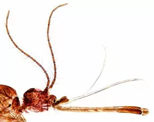
"Culex pipiens: A Fascinating Look into the World of European River Life" In this captivating artwork, titled C016 / 3451, we are presented with a close-up view of the head of a culex pipiens mosquito. This particular species is commonly known as the Northern house mosquito and can be found in various regions across Europe. As we delve deeper into the European river ecosystem, another artwork (C016 / 3450) showcases an array of insects that coexist alongside the culex pipiens. These intricate creatures play vital roles in maintaining balance within their habitat. Moving on to two pupae of the mosquito, we witness their transformation from larvae to adult form. The process is truly remarkable and highlights nature's ability to adapt and evolve. Examining these mosquitoes under different microscopes provides us with unique perspectives. In one image captured using scanning electron microscopy (SEM), we observe the detailed structure of a male culex mosquito's wing. Its delicate veins intricately patterned like fine lacework. Contrasting this, a light micrograph reveals fascinating details about female genitalia—LM allows us to explore aspects often hidden from plain sight. Additionally, another intriguing image displays a testis infected with bacteria—a reminder that even these tiny creatures face challenges in their environment. Further exploration takes us inside a mosquito ovary infected with bacteria through transmission electron microscopy (TEM). This glimpse unveils how infections impact reproductive organs—an area crucial for population dynamics and survival strategies. Returning once again to examine a culex mosquito wing through light microscopy offers yet another perspective on its intricate design—the beauty lies not only in its functionality but also in its aesthetic appeal.