
Coronary Collection
"Unveiling the Intricate Network: Exploring the Coronary System and its Impact on Heart Health" The heart


All Professionally Made to Order for Quick Shipping
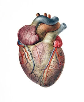
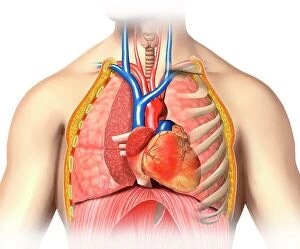
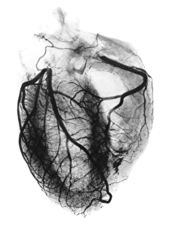
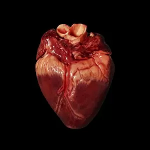
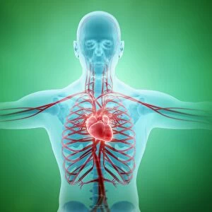
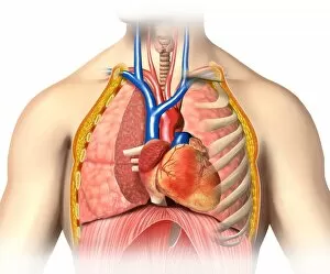
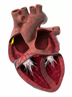
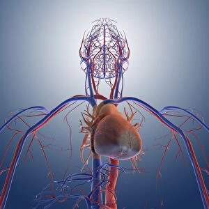
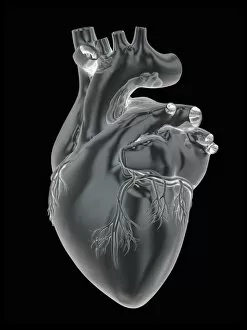
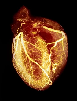
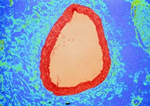
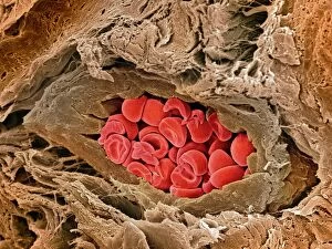
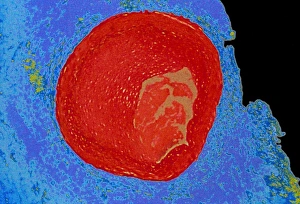
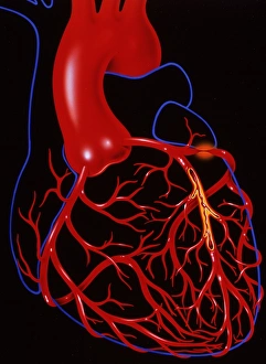
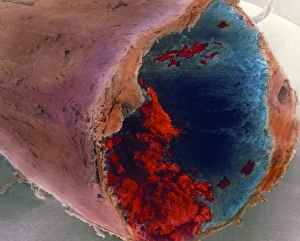
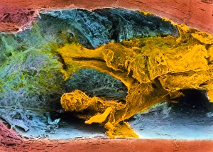
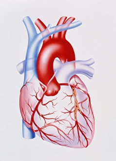
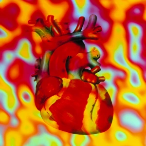
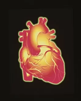
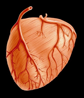
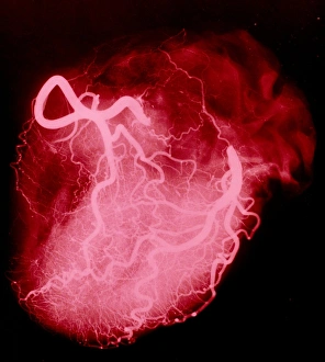
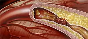
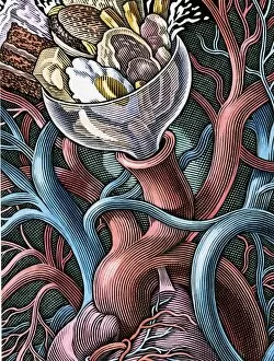
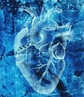
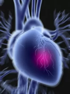
"Unveiling the Intricate Network: Exploring the Coronary System and its Impact on Heart Health" The heart, a vital organ responsible for pumping life-giving blood throughout our bodies, relies on a complex network vessels to ensure its own nourishment. With an illustration depicting a heart with coronary vessels, we delve into the intricate pathways that supply oxygen-rich blood to every nook and cranny of this remarkable muscle. A stent inside an artery serves as a reminder of medical advancements in treating coronary conditions, offering hope to those affected by blockages or narrowings within these crucial vessels. Taking us back in time, a vintage 1904 arteriogram showcases early attempts at visualizing the arteries of the heart – paving the way for modern diagnostic techniques that save countless lives today. As we examine a pig's heart closely, we gain insight into how similar our own cardiovascular system is structured – highlighting both our vulnerability and resilience when it comes to matters of the heart. Through captivating artwork featuring hearts with their intricate web vessels (F006 / 3598 & F006 / 3586), we are reminded of nature's awe-inspiring design that keeps us alive day after day. In an era where technology plays an ever-increasing role in healthcare, tablet computer illustrations depict how even something as small as this device can help raise awareness about life-threatening events like heart attacks. Combining artistry with science, ECG traces alongside MRI brain scans create visually striking compositions that symbolize the interconnectedness between cardiac health and overall well-being. These captivating artworks serve not only as educational tools but also inspire appreciation for our resilient hearts while urging us to prioritize self-care and seek timely medical attention when needed. Let us embark on this journey through stunning visuals and thought-provoking imagery – exploring all facets of the coronary system and its profound impact on our heart's health.