
Conidiophores Collection
Conidiophores, the fascinating structures of fungal spore production, are beautifully captured in this collection of micrographs. Picture No


All Professionally Made to Order for Quick Shipping
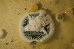
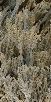
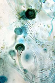
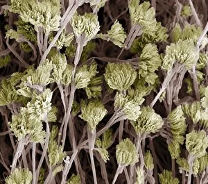
Conidiophores, the fascinating structures of fungal spore production, are beautifully captured in this collection of micrographs. Picture No. 11014633 showcases Penicillium fungal spores under scanning electron microscopy (SEM), revealing their intricate details and unique shapes. The grey mould fungus is also depicted in several light micrographs, displaying its delicate filaments and conidiophores that give rise to countless spores. The SEM images provide a closer look at the Penicillium fungus, unveiling its complex network that resemble tiny brushes or brooms. These structures play a crucial role in dispersing spores into the environment for reproduction and survival. Bread mould is another subject explored through SEM imaging, exhibiting its characteristic conidiophores that form dense clusters on the surface. These bread mould conidiophores appear like miniature trees with branches extending outward to release numerous spores.